Perrin Rosalind, Maguire Patrick, Garonna Adriano, Weidlich Georg, Bulling Shelley, Fargier-Voiron Marie, De Marco Cedric, Rossi Eleonora, Ciocca Mario, Vitolo Viviana, Mirandola Alfredo
EBAMed SA, Geneva, Switzerland.
MedDevicePharma LLC, Foster City, CA, United States.
Front Cardiovasc Med. 2022 May 4;9:849247. doi: 10.3389/fcvm.2022.849247. eCollection 2022.
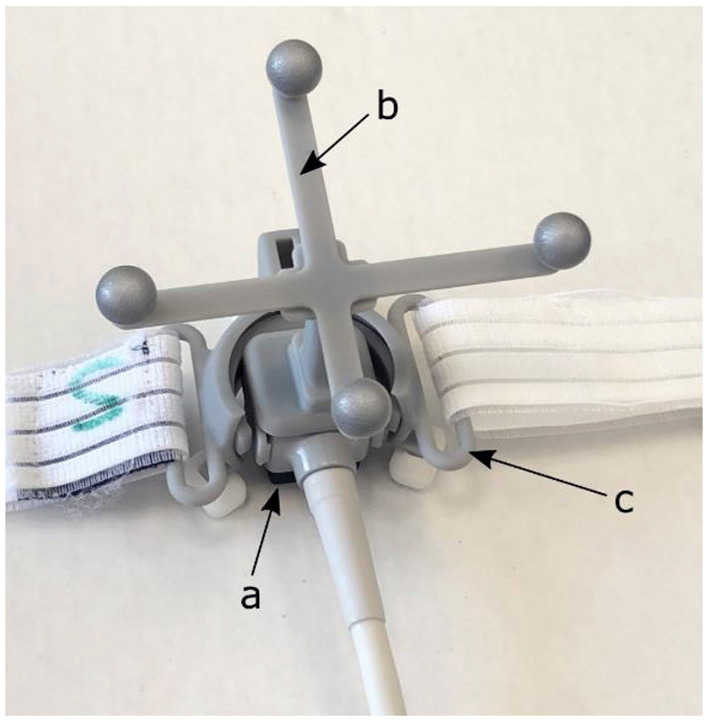
Cardiac arrhythmias, such as ventricular tachycardia, are disruptions in the normal cardiac function that originate from problems in the electrical conduction of signals inside the heart. Recently, a non-invasive treatment option based on external photon or proton beam irradiation has been used to ablate the arrhythmogenic structures. Especially in proton therapy, based on its steep dose gradient, it is crucial to monitor the motion of the heart in order to ensure that the radiation dose is delivered to the correct location. Transthoracic ultrasound imaging has the potential to provide guidance during this treatment delivery. However, it has to be noted that the presence of an ultrasound probe on the chest of the patient introduces constraints on usable beam angles for both protons and photon treatments. This case report investigates the possibility to generate a clinically acceptable proton treatment plan while the ultrasound probe is present on the chest of the patient.
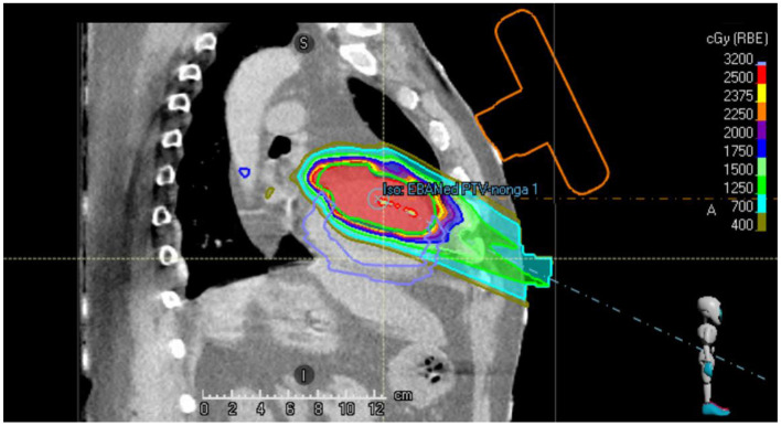
A treatment plan study was performed based on a 4D cardiac-gated computed tomography scan of a 55 year-old male patient suffering from refractory ventricular tachycardia who underwent cardiac radioablation. A proton therapy treatment plan was generated for the actual treatment target in presence of an ultrasound probe on the chest of this patient. The clinical acceptability of the generated plan was confirmed by evaluating standard target dose-volume metrics, dose to organs-at-risk and target dose conformity and homogeneity.
The generation of a clinically acceptable proton therapy treatment plan for cardiac radioablation of ventricular tachycardia could be performed in the presence of an ultrasound probe on the chest of the patient. These results establish a basis and justification for continued research and product development for ultrasound-guided cardiac radioablation.
心律失常,如室性心动过速,是正常心脏功能的紊乱,源于心脏内部信号传导问题。最近,一种基于外部光子或质子束照射的非侵入性治疗方法已被用于消融致心律失常结构。特别是在质子治疗中,基于其陡峭的剂量梯度,监测心脏运动以确保辐射剂量传递到正确位置至关重要。经胸超声成像有可能在这种治疗过程中提供指导。然而,必须注意的是,患者胸部放置超声探头会对质子和光子治疗的可用束角产生限制。本病例报告探讨了在患者胸部存在超声探头的情况下生成临床可接受的质子治疗计划的可能性。
基于一名55岁患有难治性室性心动过速并接受心脏射频消融的男性患者的4D心脏门控计算机断层扫描进行了治疗计划研究。针对该患者胸部存在超声探头的实际治疗靶点生成了质子治疗计划。通过评估标准靶区剂量体积指标、危及器官的剂量以及靶区剂量适形度和均匀性,确认了所生成计划的临床可接受性。
在患者胸部存在超声探头的情况下,可以为室性心动过速的心脏射频消融生成临床可接受的质子治疗计划。这些结果为超声引导心脏射频消融的持续研究和产品开发奠定了基础并提供了依据。