Ophthalmology Unit, University Hospital of Parma, via Gramsci 14, 43126, Parma, Italy.
Institute of Molecular and Clinical Ophthalmology Basel, Basel, Switzerland.
BMC Ophthalmol. 2022 May 23;22(1):233. doi: 10.1186/s12886-022-02430-x.
Information on the centration and tilt of iris-claw intraocular lenses (IC-IOLs) is limited. In this study, we tested the capacity of an anterior segment optical coherence tomography (AS-OCT) instrument to measure decentration and tilt of anterior and posterior IC-IOLs through an integrated software.
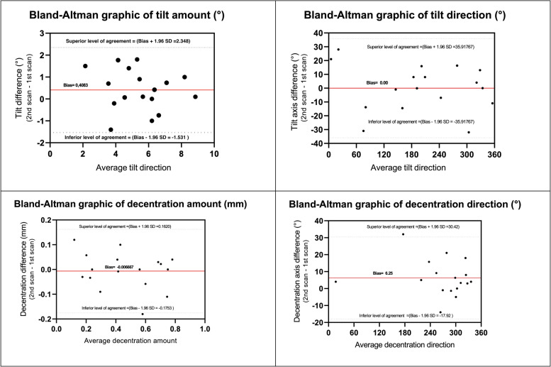
The present observational, cross-sectional study was conducted at University Eye Clinic of Parma (Parma, Italy). The CASIA2 swept-source AS-OCT (Tomey Corp.) was used to measure the tilt and decentration of posterior and anterior IC-IOLs in patients implanted at least 6 months in advance. After failure with full-automation, semi-manual IOL tracing was applied. In-the-bag (IB) contralateral IOLs, when present, were measured automatically. The Bland-Altman method was used to evaluate the agreement between repeated measurements (2 images for each study eye). The amount and direction of tilt and decentration were recorded and plotted into polar charts for evaluation.
A total of 21 patients were included: 14 with posterior and 7 with anterior IC-IOL fixation. In 17 eyes (81%), the AS-OCT provided a repeatable measurement of tilt and decentration. All contralateral eyes with IB IOL were automatically measured. The median decentration was 0.67 mm, 0.24 mm, and 0.24 mm in posterior IC-IOLs, anterior IC-IOLs, and IB IOLs group, respectively. The median tilt was 5.0°, 5.6°, and 5.6° for posterior IC-IOLs, anterior IC-IOLs, and IB IOLs, respectively. Tilt direction was mainly temporal, while decentration was inferior-temporal with posterior IC-IOLs and scattered with anterior IC-IOLs and IB IOLs.
The semi-manual tracing function of the CASIA2 AS-OCT provides repeatable and affordable measurements of the decentration and tilt of IC-IOLs in both the anterior and posterior chamber. Data from the former group were similar to the IB group.
虹膜夹型人工晶状体(IC-IOL)的偏心和倾斜信息有限。本研究通过集成软件测试了前节光学相干断层扫描(AS-OCT)仪器测量前房和后房 IC-IOL 偏心和倾斜的能力。
本观察性、横断面研究在意大利帕尔马大学眼科诊所(帕尔马)进行。使用 CASIA2 扫频源 AS-OCT(Tomey 公司)测量至少提前 6 个月植入的患者的后房型和前房型 IC-IOL 的倾斜和偏心。在全自动失败后,应用半手动 IOL 追踪。当存在时,会自动测量袋内(IB)对侧 IOL。采用 Bland-Altman 法评估重复测量(每只研究眼 2 张图像)之间的一致性。记录并绘制倾斜和偏心的量和方向到极坐标图进行评估。
共纳入 21 例患者:14 例后房型和 7 例前房型 IC-IOL 固定。在 17 只眼中(81%),AS-OCT 可重复测量倾斜和偏心。所有 IB 晶状体的对侧眼均自动测量。后房型 IC-IOL、前房型 IC-IOL 和 IB IOL 组的中位偏心量分别为 0.67mm、0.24mm 和 0.24mm。后房型 IC-IOL、前房型 IC-IOL 和 IB IOL 的中位倾斜度分别为 5.0°、5.6°和 5.6°。倾斜方向主要为颞侧,而偏心则以后侧 IC-IOL 为主,前房型 IC-IOL 和 IB IOL 则为分散性。
CASIA2 AS-OCT 的半手动追踪功能可重复、经济地测量前房和后房 IC-IOL 的偏心和倾斜。前组数据与 IB 组相似。