Kimura Shuhei, Morizane Yuki, Shiode Yusuke, Hirano Masayuki, Doi Shinichiro, Toshima Shinji, Fujiwara Atsushi, Shiraga Fumio
Department of Ophthalmology, Okayama University Graduate School of Medicine, Dentistry and Pharmaceutical Sciences, Okayama, Japan.
PLoS One. 2017 Sep 1;12(9):e0184066. doi: 10.1371/journal.pone.0184066. eCollection 2017.
To investigate the tilt and decentration of the crystalline lens and the intraocular lens (IOL) relative to the corneal topographic axis using anterior segment ocular coherence tomography (AS-OCT).
A sample set of 100 eyes from 49 subjects (41 eyes with crystalline lenses and 59 eyes with IOLs) were imaged using second generation AS-OCT (CASIA2, TOMEY) in June and July 2016 at Okayama University. Both mydriatic and non-mydriatic images were obtained, and the tilt and decentration of the crystalline lens and the IOL were quantified. The effects of pupil dilation on measurements were also assessed.
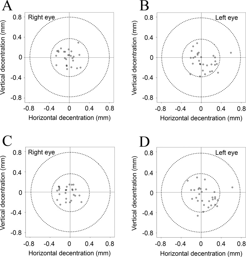
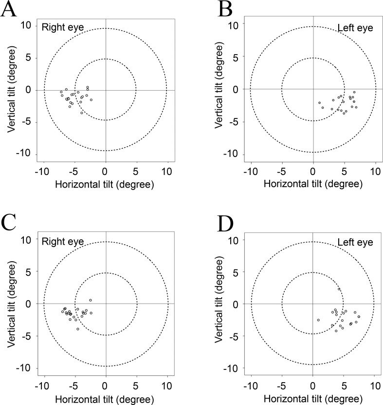
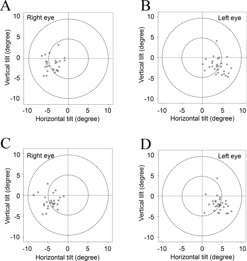
The crystalline lens showed an average tilt of 5.15° towards the inferotemporal direction relative to the corneal topographic axis under non-mydriatic conditions and 5.25° under mydriatic conditions. Additionally, an average decentration of 0.11 mm towards the temporal direction was observed under non-mydriatic conditions and 0.08 mm under mydriatic conditions. The average tilt for the IOL was 4.31° towards the inferotemporal direction relative to the corneal topographic axis under non-mydriatic conditions and 4.65° in the same direction under mydriatic conditions. The average decentration was 0.05 mm towards the temporal direction under non-mydriatic conditions and 0.08 mm in the same direction under mydriatic conditions. A strong correlation was found between the average tilt and decentration values of the crystalline lens and the IOL under both non-mydriatic and mydriatic conditions (all Spearman correlation coefficients, r ≥ 0.800; all P < 0.001).
When measured using second generation AS-OCT, both the crystalline lens and the IOL showed an average tilt of 4-6° toward the inferotemporal direction relative to the corneal topographic axis and an average decentration of less than 0.12 mm towards the temporal direction. These results were not influenced by pupil dilation and they showed good repeatability.
使用眼前节光学相干断层扫描(AS-OCT)研究晶状体和人工晶状体(IOL)相对于角膜地形图轴的倾斜和偏心情况。
2016年6月和7月在冈山大学使用第二代AS-OCT(CASIA2,TOMEY)对49名受试者的100只眼(41只眼有晶状体,59只眼有人工晶状体)进行成像。获取散瞳和未散瞳图像,并对晶状体和人工晶状体的倾斜和偏心进行量化。还评估了瞳孔散大对测量的影响。
在未散瞳条件下,晶状体相对于角膜地形图轴平均向颞下方向倾斜5.15°,散瞳条件下为5.25°。此外,未散瞳条件下平均向颞侧偏心0.11 mm,散瞳条件下为0.08 mm。人工晶状体在未散瞳条件下相对于角膜地形图轴平均向颞下方向倾斜4.31°,散瞳条件下在同一方向为4.65°。未散瞳条件下平均向颞侧偏心0.05 mm,散瞳条件下在同一方向为0.08 mm。在未散瞳和散瞳条件下,晶状体和人工晶状体的平均倾斜和偏心值之间均发现强相关性(所有Spearman相关系数,r≥0.800;所有P<0.001)。
使用第二代AS-OCT测量时,晶状体和人工晶状体相对于角膜地形图轴均平均向颞下方向倾斜4-6°,平均向颞侧偏心小于0.12 mm。这些结果不受瞳孔散大影响,且具有良好的重复性。