Translational Research Program in Pediatric Orthopaedics, Division of Orthopaedic Surgery, Department of Surgery, The Children's Hospital of Philadelphia, Philadelphia, PA 19104, USA.
Department of Bioengineering, Temple University, Philadelphia, PA 19122, USA.
Biol Open. 2022 Jun 15;11(6). doi: 10.1242/bio.059381.
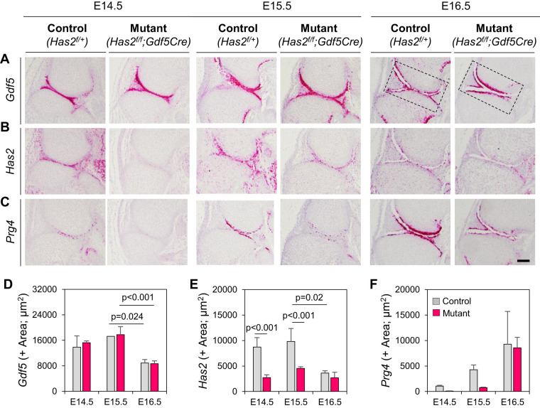
The synovial cavity and its fluid are essential for joint function and lubrication, but their developmental biology remains largely obscure. Here, we analyzed E12.5 to E18.5 mouse embryo hindlimbs and discovered that cavitation initiates around E15.0 with emergence of multiple, discrete, µm-wide tissue discontinuities we term microcavities in interzone, evolving into a single joint-wide cavity within 12 h in knees and within 72-84 h in interphalangeal joints. The microcavities were circumscribed by cells as revealed by mTmG imaging and exhibited a carbohydrate and protein content based on infrared spectral imaging at micro and nanoscale. Accounting for differing cavitation kinetics, we found that the growing femur and tibia anlagen progressively flexed at the knee over time, with peak angulation around E15.5 exactly when the full knee cavity consolidated; however, interphalangeal joint geometry changed minimally over time. Indeed, cavitating knee interzone cells were elongated along the flexion angle axis and displayed oblong nuclei, but these traits were marginal in interphalangeal cells. Conditional Gdf5Cre-driven ablation of Has2 - responsible for production of the joint fluid component hyaluronic acid (HA) - delayed the cavitation process. Our data reveal that cavitation is a stepwise process, brought about by sequential action of microcavities, skeletal flexion and elongation, and HA accumulation. This article has an associated First Person interview with the first author of the paper.
滑液腔及其液体对于关节的功能和润滑至关重要,但它们的发育生物学仍然很大程度上不为人知。在这里,我们分析了 E12.5 至 E18.5 天的小鼠胚胎后肢,发现 E15.0 左右开始出现空化,在间区出现多个离散的、宽数微米的组织不连续性,我们称之为微腔,在 12 小时内在膝关节内发展成单个关节宽的腔,在 72-84 小时内在指间关节内发展成单个关节宽的腔。微腔被细胞包围,这一点通过 mTmG 成像显示出来,并且在微纳尺度上基于红外光谱成像显示出碳水化合物和蛋白质含量。考虑到不同的空化动力学,我们发现不断生长的股骨和胫骨原基随着时间的推移在膝关节处逐渐弯曲,在 E15.5 左右时最大弯曲角度出现,此时整个膝关节腔完全融合;然而,指间关节的几何形状随时间变化很小。事实上,空化的膝关节间区细胞沿弯曲角度轴拉长,并显示出长形核,但这些特征在指间关节细胞中微不足道。条件性 Gdf5Cre 驱动的 Has2 消融 - 负责关节液成分透明质酸 (HA) 的产生 - 延迟了空化过程。我们的数据显示,空化是一个逐步的过程,是由微腔、骨骼弯曲和伸长以及 HA 积累的顺序作用引起的。本文附有该论文第一作者的相关第一人称采访。