• 文献检索
  • 文档翻译
  • 深度研究
  • 学术资讯
  • Suppr Zotero 插件Zotero 插件
  • 邀请有礼
  • 套餐&价格
  • 历史记录
应用&插件
Suppr Zotero 插件Zotero 插件浏览器插件Mac 客户端Windows 客户端微信小程序
定价
高级版会员购买积分包购买API积分包
服务
文献检索文档翻译深度研究API 文档MCP 服务
关于我们
关于 Suppr公司介绍联系我们用户协议隐私条款
关注我们

Suppr 超能文献

核心技术专利:CN118964589B侵权必究
粤ICP备2023148730 号-1Suppr @ 2026

文献检索

告别复杂PubMed语法,用中文像聊天一样搜索,搜遍4000万医学文献。AI智能推荐,让科研检索更轻松。

立即免费搜索

文件翻译

保留排版,准确专业,支持PDF/Word/PPT等文件格式,支持 12+语言互译。

免费翻译文档

深度研究

AI帮你快速写综述,25分钟生成高质量综述,智能提取关键信息,辅助科研写作。

立即免费体验

利用活性微柱阵列测量细胞骨架的力学波动和流变学性质。

Measuring Cytoskeletal Mechanical Fluctuations and Rheology with Active Micropost Arrays.

机构信息

Department of Physics and Astronomy, Johns Hopkins University, Baltimore, Maryland.

Current address: Department of Biomedical Engineering, University of North Carolina, Chapel Hill, North Carolina.

出版信息

Curr Protoc. 2022 May;2(5):e433. doi: 10.1002/cpz1.433.

DOI:10.1002/cpz1.433
PMID:35612274
原文链接:https://pmc.ncbi.nlm.nih.gov/articles/PMC9321978/
Abstract

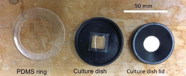
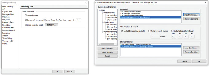
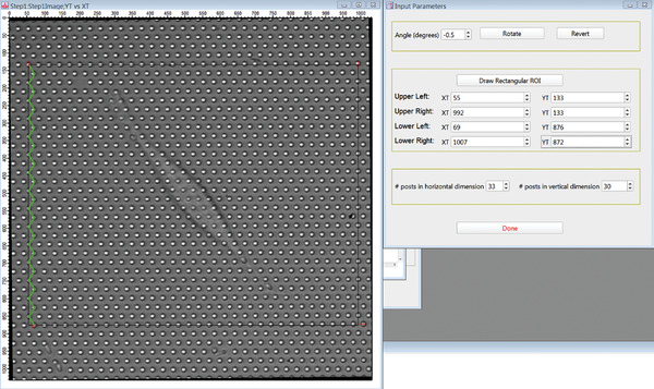
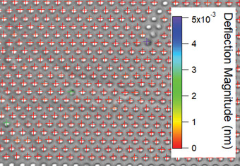
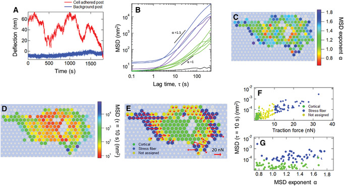
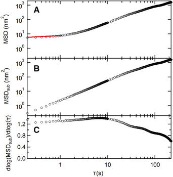
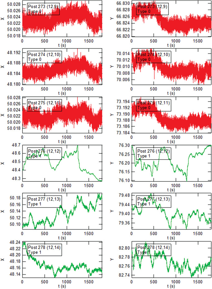
The dynamics of the cellular actomyosin cytoskeleton are crucial to many aspects of cellular function. Here, we describe techniques that employ active micropost array detectors (AMPADs) to measure cytoskeletal rheology and mechanical force fluctuations. The AMPADS are arrays of flexible poly(dimethylsiloxane) (PDMS) microposts with magnetic nanowires embedded in a subset of microposts to enable actuation of those posts via an externally applied magnetic field. Techniques are described to track the magnetic microposts' motion with nanometer precision at up to 100 video frames per second to measure the local cellular rheology at well-defined positions. Application of these high-precision tracking techniques to the full array of microposts in contact with a cell also enables mapping of the cytoskeletal mechanical fluctuation dynamics with high spatial and temporal resolution. This article describes (1) the fabrication of magnetic micropost arrays, (2) measurement protocols for both local rheology and cytoskeletal force fluctuation mapping, and (3) special-purpose software routines to reduce and analyze these data. © 2022 The Authors. Current Protocols published by Wiley Periodicals LLC. Basic Protocol 1: Fabrication of magnetic micropost arrays Basic Protocol 2: Data acquisition for cellular force fluctuations on non-magnetic micropost arrays Basic Protocol 3: Data acquisition for local cellular rheology measurements with magnetic microposts Basic Protocol 4: Data reduction: determining microposts' motion Basic Protocol 5: Data analysis: determining local rheology from magnetic microposts Basic Protocol 6: Data analysis for force fluctuation measurements Support Protocol 1: Fabrication of magnetic Ni nanowires by electrodeposition Support Protocol 2: Configuring Streampix for magnetic rheology measurements.

摘要

细胞肌动球蛋白细胞骨架的动力学对细胞功能的许多方面都至关重要。在这里,我们描述了使用主动微柱阵列探测器(AMPAD)来测量细胞骨架流变学和机械力波动的技术。AMPADS 是由柔性聚二甲基硅氧烷(PDMS)微柱阵列组成,其中一些微柱中嵌入了磁性纳米线,以便通过外部施加的磁场来驱动这些微柱的运动。本文介绍了以纳米级精度跟踪磁性微柱运动的技术,其速度高达每秒 100 个视频帧,以测量在明确定义位置处的局部细胞流变学。将这些高精度跟踪技术应用于与细胞接触的整个微柱阵列,还可以以高时空分辨率绘制细胞骨架机械波动动力学图。本文描述了(1)磁性微柱阵列的制作,(2)局部流变学和细胞骨架力波动映射的测量方案,以及(3)用于减少和分析这些数据的专用软件例程。 © 2022 作者。Wiley Periodicals LLC 出版的《当代协议》。 基本方案 1:磁性微柱阵列的制作 基本方案 2:非磁性微柱阵列上细胞力波动的数据采集 基本方案 3:使用磁性微柱进行局部细胞流变学测量的数据采集 基本方案 4:数据减少:确定微柱的运动 基本方案 5:数据分析:从磁性微柱确定局部流变学 基本方案 6:力波动测量数据分析 支持方案 1:通过电沉积制备磁性 Ni 纳米线 支持方案 2:为磁性流变学测量配置 Streampix。

https://cdn.ncbi.nlm.nih.gov/pmc/blobs/b69f/9321978/7b8f2cbc37f0/CPZ1-2-0-g021.jpg
https://cdn.ncbi.nlm.nih.gov/pmc/blobs/b69f/9321978/5a0fb1bbc9df/CPZ1-2-0-g007.jpg
https://cdn.ncbi.nlm.nih.gov/pmc/blobs/b69f/9321978/5f8a292e36ad/CPZ1-2-0-g037.jpg
https://cdn.ncbi.nlm.nih.gov/pmc/blobs/b69f/9321978/489da190d658/CPZ1-2-0-g031.jpg
https://cdn.ncbi.nlm.nih.gov/pmc/blobs/b69f/9321978/5d664b819c58/CPZ1-2-0-g014.jpg
https://cdn.ncbi.nlm.nih.gov/pmc/blobs/b69f/9321978/9c810cf98883/CPZ1-2-0-g012.jpg
https://cdn.ncbi.nlm.nih.gov/pmc/blobs/b69f/9321978/14d4ad50aa58/CPZ1-2-0-g023.jpg
https://cdn.ncbi.nlm.nih.gov/pmc/blobs/b69f/9321978/6ec5f0c63d1f/CPZ1-2-0-g025.jpg
https://cdn.ncbi.nlm.nih.gov/pmc/blobs/b69f/9321978/6d775d8133c6/CPZ1-2-0-g004.jpg
https://cdn.ncbi.nlm.nih.gov/pmc/blobs/b69f/9321978/8b8f325f7622/CPZ1-2-0-g003.jpg
https://cdn.ncbi.nlm.nih.gov/pmc/blobs/b69f/9321978/ed5847eab355/CPZ1-2-0-g013.jpg
https://cdn.ncbi.nlm.nih.gov/pmc/blobs/b69f/9321978/334703fbf28c/CPZ1-2-0-g015.jpg
https://cdn.ncbi.nlm.nih.gov/pmc/blobs/b69f/9321978/ee03619a2be1/CPZ1-2-0-g024.jpg
https://cdn.ncbi.nlm.nih.gov/pmc/blobs/b69f/9321978/59b9d378452b/CPZ1-2-0-g022.jpg
https://cdn.ncbi.nlm.nih.gov/pmc/blobs/b69f/9321978/20febe6211b5/CPZ1-2-0-g008.jpg
https://cdn.ncbi.nlm.nih.gov/pmc/blobs/b69f/9321978/30bdd39d596d/CPZ1-2-0-g002.jpg
https://cdn.ncbi.nlm.nih.gov/pmc/blobs/b69f/9321978/2b19be33f6dd/CPZ1-2-0-g030.jpg
https://cdn.ncbi.nlm.nih.gov/pmc/blobs/b69f/9321978/c4d7818c98b8/CPZ1-2-0-g036.jpg
https://cdn.ncbi.nlm.nih.gov/pmc/blobs/b69f/9321978/4626f8b0e6e5/CPZ1-2-0-g016.jpg
https://cdn.ncbi.nlm.nih.gov/pmc/blobs/b69f/9321978/f158c6c1df78/CPZ1-2-0-g011.jpg
https://cdn.ncbi.nlm.nih.gov/pmc/blobs/b69f/9321978/a6d20b597204/CPZ1-2-0-g032.jpg
https://cdn.ncbi.nlm.nih.gov/pmc/blobs/b69f/9321978/fdced9682ef5/CPZ1-2-0-g034.jpg
https://cdn.ncbi.nlm.nih.gov/pmc/blobs/b69f/9321978/d4c23b477abb/CPZ1-2-0-g006.jpg
https://cdn.ncbi.nlm.nih.gov/pmc/blobs/b69f/9321978/577a46c4b0cc/CPZ1-2-0-g039.jpg
https://cdn.ncbi.nlm.nih.gov/pmc/blobs/b69f/9321978/1e7f0ac195b5/CPZ1-2-0-g027.jpg
https://cdn.ncbi.nlm.nih.gov/pmc/blobs/b69f/9321978/fb677674f25d/CPZ1-2-0-g019.jpg
https://cdn.ncbi.nlm.nih.gov/pmc/blobs/b69f/9321978/866b32b5eb3f/CPZ1-2-0-g010.jpg
https://cdn.ncbi.nlm.nih.gov/pmc/blobs/b69f/9321978/a14bc42b1c47/CPZ1-2-0-g018.jpg
https://cdn.ncbi.nlm.nih.gov/pmc/blobs/b69f/9321978/63be3d04140e/CPZ1-2-0-g038.jpg
https://cdn.ncbi.nlm.nih.gov/pmc/blobs/b69f/9321978/221d83c29530/CPZ1-2-0-g029.jpg
https://cdn.ncbi.nlm.nih.gov/pmc/blobs/b69f/9321978/d81ca4f953d6/CPZ1-2-0-g020.jpg
https://cdn.ncbi.nlm.nih.gov/pmc/blobs/b69f/9321978/0e2f8c3ebf0d/CPZ1-2-0-g028.jpg
https://cdn.ncbi.nlm.nih.gov/pmc/blobs/b69f/9321978/6a6e8f20329c/CPZ1-2-0-g017.jpg
https://cdn.ncbi.nlm.nih.gov/pmc/blobs/b69f/9321978/ad89734e6fbc/CPZ1-2-0-g009.jpg
https://cdn.ncbi.nlm.nih.gov/pmc/blobs/b69f/9321978/3978ca32199e/CPZ1-2-0-g035.jpg
https://cdn.ncbi.nlm.nih.gov/pmc/blobs/b69f/9321978/131d92af7e03/CPZ1-2-0-g033.jpg
https://cdn.ncbi.nlm.nih.gov/pmc/blobs/b69f/9321978/f2831a57e15b/CPZ1-2-0-g001.jpg
https://cdn.ncbi.nlm.nih.gov/pmc/blobs/b69f/9321978/54f09b8c6d35/CPZ1-2-0-g005.jpg
https://cdn.ncbi.nlm.nih.gov/pmc/blobs/b69f/9321978/4043178cf3fe/CPZ1-2-0-g026.jpg
https://cdn.ncbi.nlm.nih.gov/pmc/blobs/b69f/9321978/7b8f2cbc37f0/CPZ1-2-0-g021.jpg

相似文献

1
Measuring Cytoskeletal Mechanical Fluctuations and Rheology with Active Micropost Arrays.利用活性微柱阵列测量细胞骨架的力学波动和流变学性质。
Curr Protoc. 2022 May;2(5):e433. doi: 10.1002/cpz1.433.
2
Easy and Accurate Mechano-profiling on Micropost Arrays.微柱阵列上简便且精确的机械特性分析
J Vis Exp. 2015 Nov 17(105):53350. doi: 10.3791/53350.
3
Magnetic microposts for mechanical stimulation of biological cells: fabrication, characterization, and analysis.用于对生物细胞进行机械刺激的磁性微柱:制造、表征与分析
Rev Sci Instrum. 2008 Apr;79(4):044302. doi: 10.1063/1.2906228.
4
PDMS Elastic Micropost Arrays for Studying Vascular Smooth Muscle Cells.用于研究血管平滑肌细胞的聚二甲基硅氧烷弹性微柱阵列
Sens Actuators B Chem. 2013 Nov 1;188:1055-1063. doi: 10.1016/j.snb.2013.08.018.
5
Magnetic microposts as an approach to apply forces to living cells.磁性微柱作为一种向活细胞施加力的方法。
Proc Natl Acad Sci U S A. 2007 Sep 11;104(37):14553-8. doi: 10.1073/pnas.0611613104. Epub 2007 Sep 5.
6
Probing cellular traction forces with magnetic nanowires and microfabricated force sensor arrays.用磁纳米线和微加工力传感器阵列探测细胞牵引力。
Nanotechnology. 2012 Feb 24;23(7):075101. doi: 10.1088/0957-4484/23/7/075101. Epub 2012 Jan 20.
7
Hydrogel Micropost Arrays with Single Post Tunability to Study Cell Volume and Mechanotransduction.水凝胶微柱阵列,具有单个微柱可调性,用于研究细胞体积和机械转导。
Adv Biosyst. 2020 Nov;4(11):e2000012. doi: 10.1002/adbi.202000012. Epub 2020 Oct 14.
8
Live-cell subcellular measurement of cell stiffness using a microengineered stretchable micropost array membrane.使用微工程可拉伸微柱阵列膜对活细胞的亚细胞进行细胞硬度的测量。
Integr Biol (Camb). 2012 Oct;4(10):1289-98. doi: 10.1039/c2ib20134h.
9
For whom the cells pull: Hydrogel and micropost devices for measuring traction forces.细胞为谁而牵拉:用于测量牵拉力的水凝胶和微柱装置。
Methods. 2016 Feb 1;94:51-64. doi: 10.1016/j.ymeth.2015.08.005. Epub 2015 Aug 8.
10
Micro-elastometry on whole blood clots using actuated surface-attached posts (ASAPs).使用驱动式表面附着柱(ASAPs)对全血凝块进行微弹性测量。
Lab Chip. 2015 Mar 7;15(5):1385-93. doi: 10.1039/c4lc01478b.

引用本文的文献

1
All-Covalent Nuclease-Resistant and Hydrogel-Tethered DNA Hairpin Probes Map pN Cell Traction Forces.全共价核酸酶抗性和水凝胶连接的 DNA 发夹探针绘制 pN 细胞牵引力图谱。
ACS Appl Mater Interfaces. 2023 Jul 19;15(28):33362-33372. doi: 10.1021/acsami.3c04826. Epub 2023 Jul 6.

本文引用的文献

1
Pervasive cytoquakes in the actomyosin cortex across cell types and substrate stiffness.细胞类型和基质硬度广泛存在于细胞皮层中的肌动球蛋白细胞震颤。
Integr Biol (Camb). 2021 Dec 15;13(10):246-257. doi: 10.1093/intbio/zyab017.
2
Understanding cytoskeletal avalanches using mechanical stability analysis.利用力学稳定性分析理解细胞骨架崩塌。
Proc Natl Acad Sci U S A. 2021 Oct 12;118(41). doi: 10.1073/pnas.2110239118.
3
Large and reversible myosin-dependent forces in rigidity sensing.刚性感知中依赖肌球蛋白的大且可逆的力
Nat Phys. 2019 Jul;15(7):689-695. doi: 10.1038/s41567-019-0477-9. Epub 2019 Apr 8.
4
Innovative Tools for Mechanobiology: Unraveling Outside-In and Inside-Out Mechanotransduction.力学生物学的创新工具:解析由外向内和由内向外的机械转导
Front Bioeng Biotechnol. 2019 Jul 16;7:162. doi: 10.3389/fbioe.2019.00162. eCollection 2019.
5
Dissecting fat-tailed fluctuations in the cytoskeleton with active micropost arrays.利用活性微柱阵列剖析细胞骨架中的胖尾波动。
Proc Natl Acad Sci U S A. 2019 Jul 9;116(28):13839-13846. doi: 10.1073/pnas.1900963116. Epub 2019 Jun 25.
6
Cellular Mechanotransduction: From Tension to Function.细胞机械转导:从张力到功能
Front Physiol. 2018 Jul 5;9:824. doi: 10.3389/fphys.2018.00824. eCollection 2018.
7
Fabrication and Mechanical Properties Measurements of 3D Microtissues for the Study of Cell-Matrix Interactions.用于细胞-基质相互作用研究的3D微组织的制备与力学性能测量
Methods Mol Biol. 2018;1722:303-328. doi: 10.1007/978-1-4939-7553-2_18.
8
Atomic Force Microscopy in Characterizing Cell Mechanics for Biomedical Applications: A Review.用于生物医学应用的细胞力学特性表征中的原子力显微镜:综述
IEEE Trans Nanobioscience. 2017 Sep;16(6):523-540. doi: 10.1109/TNB.2017.2714462. Epub 2017 Jun 9.
9
Non-equilibrium cytoquake dynamics in cytoskeletal remodeling and stabilization.细胞骨架重构和稳定中的非平衡细胞颤动动力学。
Soft Matter. 2016 Oct 19;12(41):8506-8511. doi: 10.1039/c6sm01041e.
10
Tropomyosin controls sarcomere-like contractions for rigidity sensing and suppressing growth on soft matrices.原肌球蛋白控制肌节样收缩,以实现刚性感知并抑制在柔软基质上的生长。
Nat Cell Biol. 2016 Jan;18(1):33-42. doi: 10.1038/ncb3277. Epub 2015 Nov 30.