Yuan Bei, Han Songbo, Yang Shaomin, Zhang Lihua, Jiang Liang, Wei Feng, Yuan Huishu, Liu Xiaoguang, Liu Zhongjun
Department of Orthopaedics, Peking University Third Hospital, No. 49 North Garden Road, Haidian District, Beijing, 100191, China.
Department of Orthopaedics, Beijing Tsinghua Changgung Hospital, School of Clinical Medicine, Tsinghua University, Beijing, 102218, China.
Insights Imaging. 2022 May 26;13(1):93. doi: 10.1186/s13244-022-01226-3.
To analyze the radiologic and clinical changes after denosumab treatment in patients with giant cell tumors (GCTs) in the mobile spine.
Clinical data and images by computed tomography and magnetic resonance imaging at a single center were retrospectively reviewed before and after denosumab treatment.
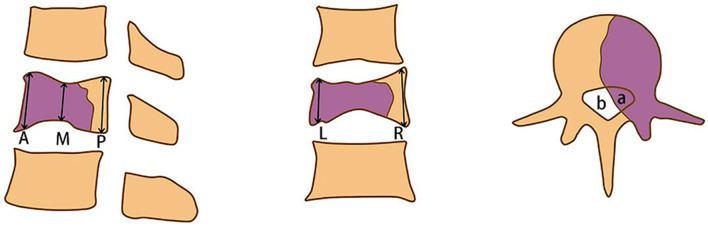
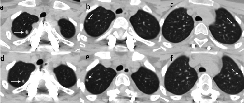
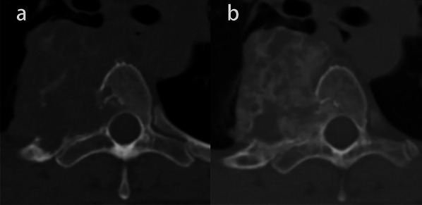
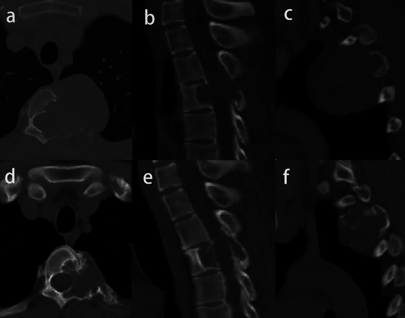
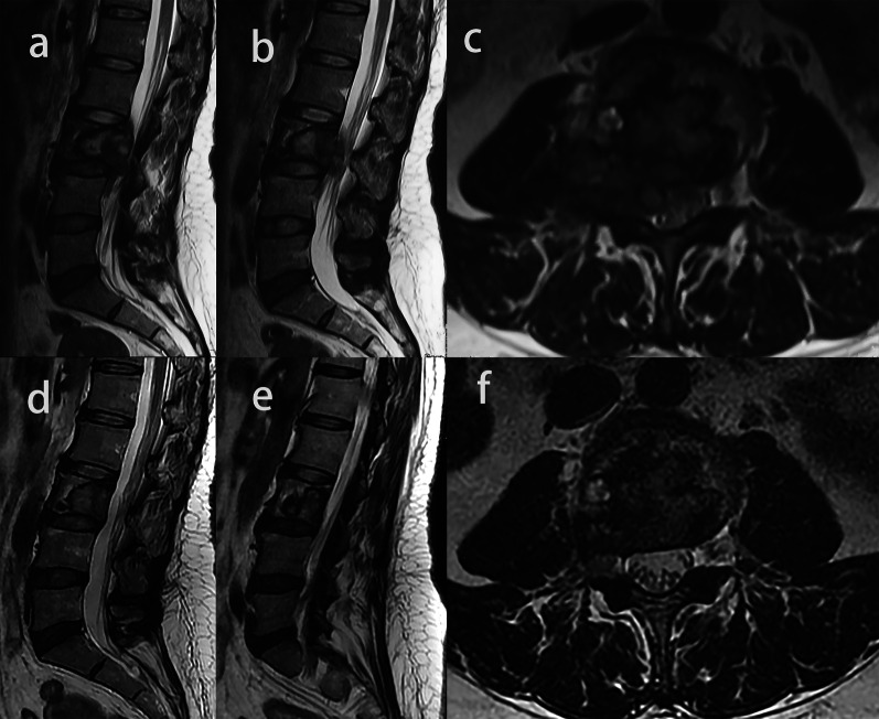
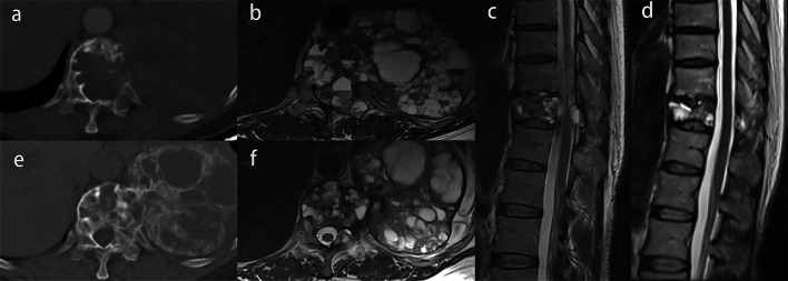
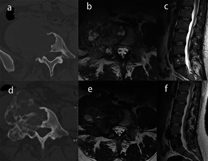
Pre- and post-treatment data from 24 patients were evaluated. On imaging, marginal ossification and/or bone formation was observed in 22 patients (91.7%). The median maximum diameter of the GCT reduced from 52.5 to 48.2 mm (p < 0.001), and the mean proportion of tumor to spinal canal area decreased from 36.8 to 18.5% (p < 0.001). Out of six patients with compression, three patients (50%) showed no compression after treatment. The signal intensity (SI) ratio between the solid part of the tumor and the normal spinal cord on T2-weighted MR images was 0.77 ± 0.22 and decreased to 0.58 ± 0.22 (p = 0.001). On clinical symptoms, the mean visual analog scale scores were reduced from 5.3 to 2.0 (p < 0.001) and the Karnofsky Performance Scale scores increased from a median of 65 to 80 (p < 0.001). Post-treatment, performance scores improved in eight patients (33.3%) (p = 0.003), and the neurological function of four patients improved according to Frankel grade (p = 0.046).
Bone formation, tumor reduction, regression of epidural lesion and the decrease in SI ratio on T2-weighted image should be considered as the effectiveness of denosumab in the treatment of spinal GCT. In clinical application, denosumab can relieve pain, improve neurological function, and improve the quality of life of spinal GCT patients.
分析地诺单抗治疗活动期脊柱骨巨细胞瘤(GCT)患者后的影像学和临床变化。
回顾性分析单中心地诺单抗治疗前后的临床资料以及计算机断层扫描和磁共振成像图像。
评估了24例患者治疗前后的数据。影像学上,22例患者(91.7%)观察到边缘骨化和/或骨形成。GCT的最大直径中位数从52.5 mm降至48.2 mm(p<0.001),肿瘤与椎管面积的平均比例从36.8%降至18.5%(p<0.001)。6例有压迫症状的患者中,3例(50%)治疗后无压迫症状。肿瘤实体部分与正常脊髓在T2加权磁共振图像上的信号强度(SI)比值为0.77±0.22,降至0.58±0.22(p=0.001)。临床症状方面,视觉模拟量表平均评分从5.3降至2.0(p<0.001),卡氏功能状态评分中位数从65提高到80(p<0.001)。治疗后,8例患者(33.3%)的功能评分改善(p=0.003),4例患者的神经功能按Frankel分级改善(p=0.046)。
骨形成、肿瘤缩小、硬膜外病变消退以及T2加权图像上SI比值降低应被视为地诺单抗治疗脊柱GCT的疗效。在临床应用中,地诺单抗可缓解疼痛、改善神经功能并提高脊柱GCT患者的生活质量。