Creze Maud, Ghaouche Jessica, Missenard Gilles, Lazure Thierry, Cluzel Guillaume, Devilder Matthieu, Briand Sylvain, Soubeyrand Marc, Meyrignac Olivier, Carlier Robert-Yves, Court Charles, Bouthors Charlie
Department of Radiology, Assistance Publique des Hôpitaux de Paris, GH Université Paris- Saclay, DMU Smart Imaging, Bicêtre Teaching Hospital, Le Kremlin-Bicêtre, France.
BioMaps, Université Paris-Saclay, Hôpital Kremlin-Bicêtre, 78 rue du Général Leclerc, 94270, Le Kremlin-Bicêtre, France.
Insights Imaging. 2023 Jul 19;14(1):128. doi: 10.1186/s13244-023-01462-1.
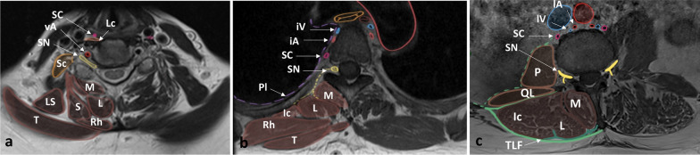
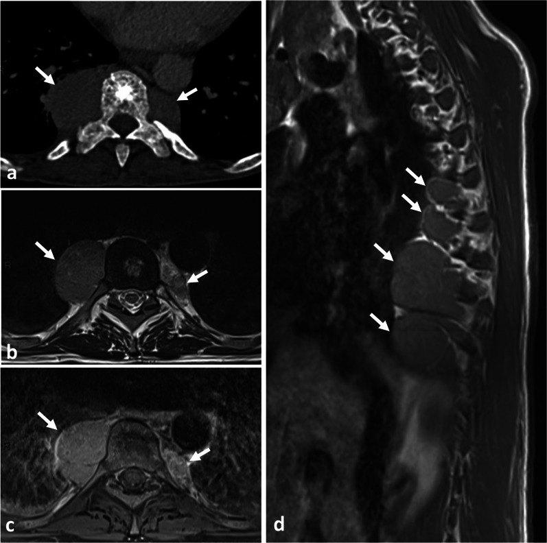
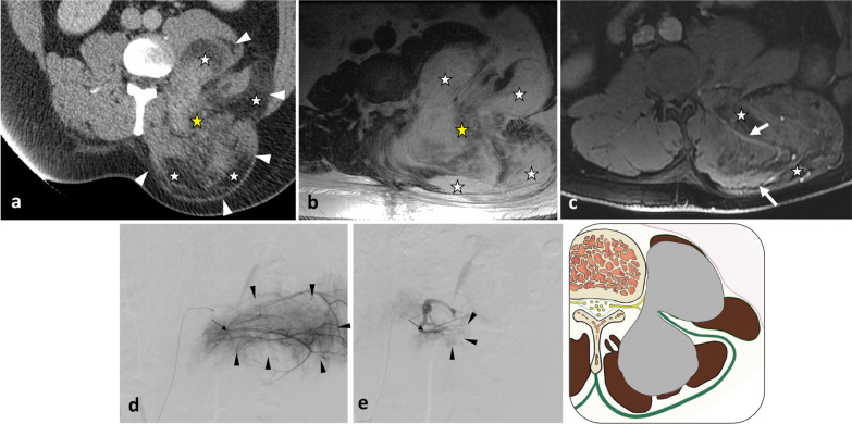
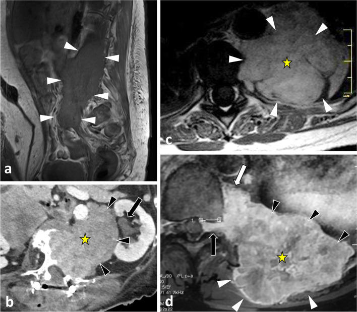
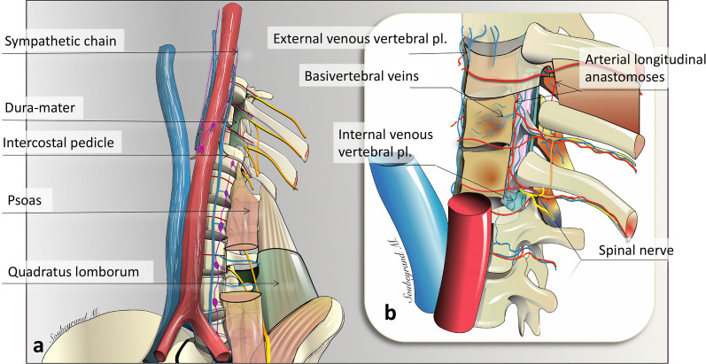
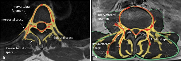
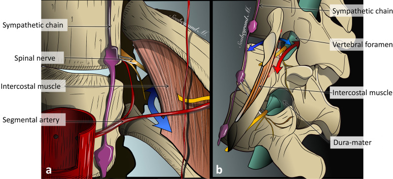
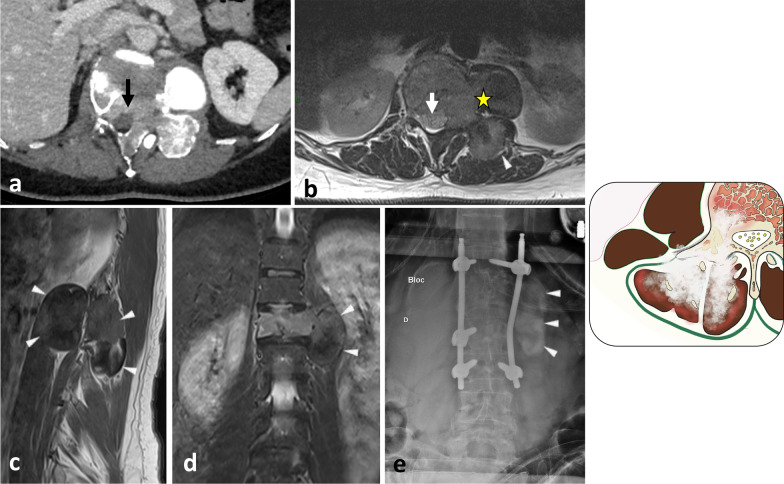
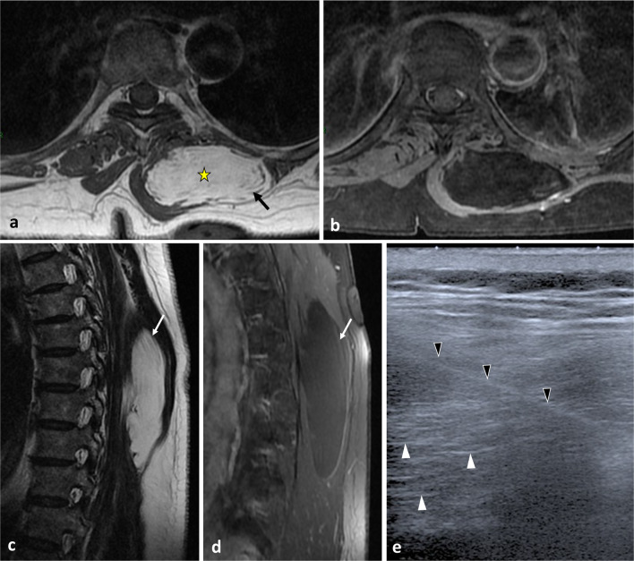
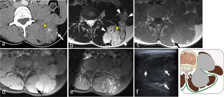
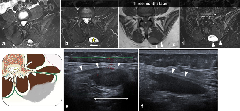
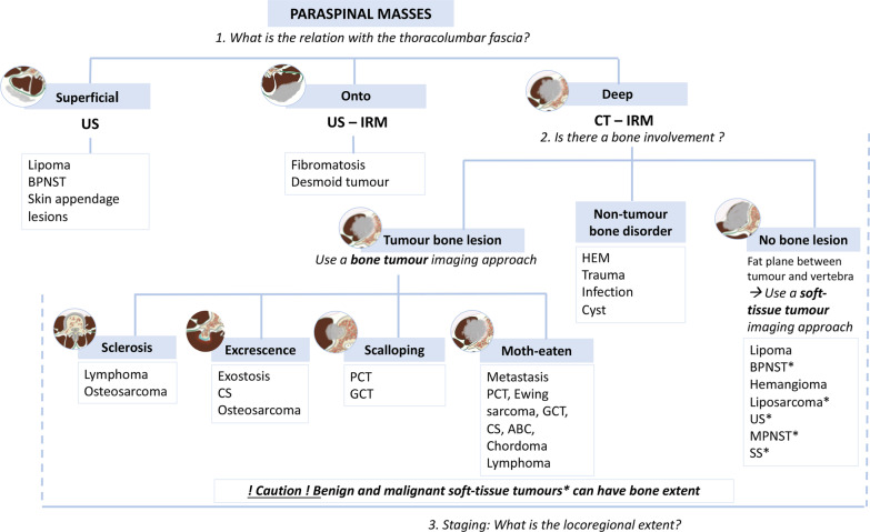
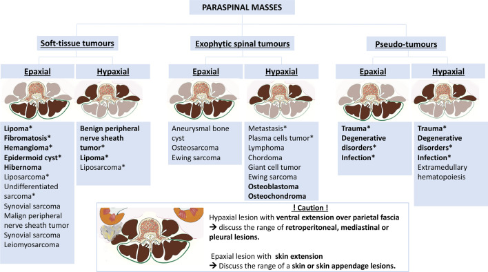
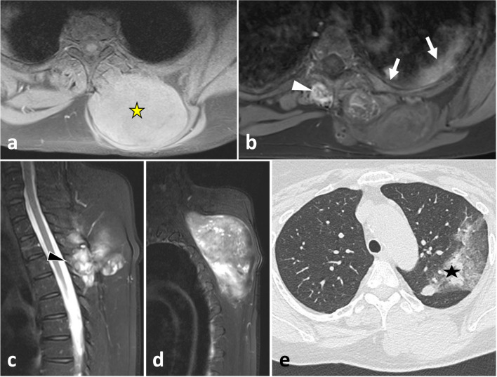
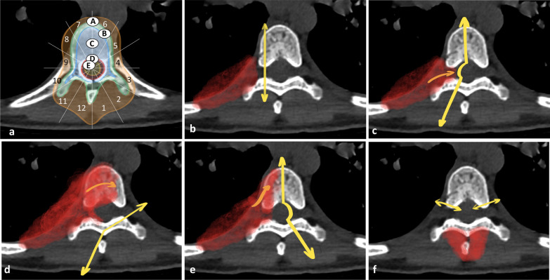
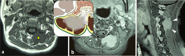
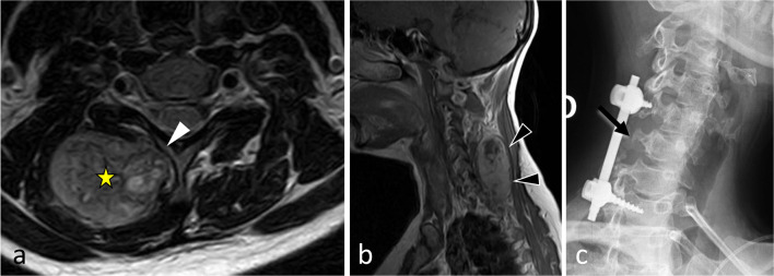
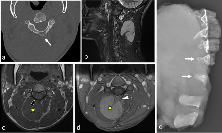
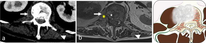
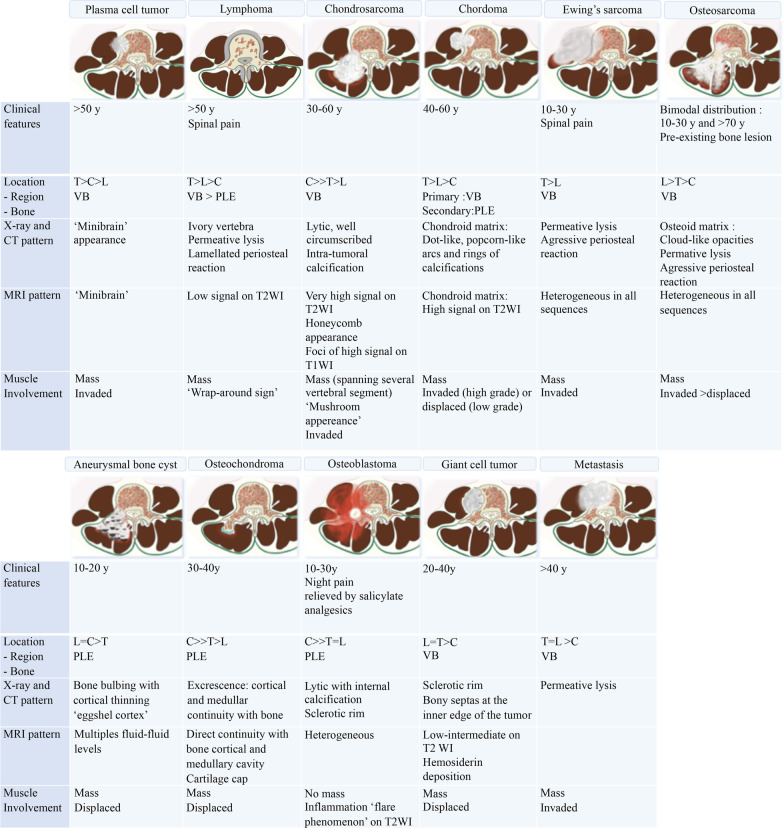
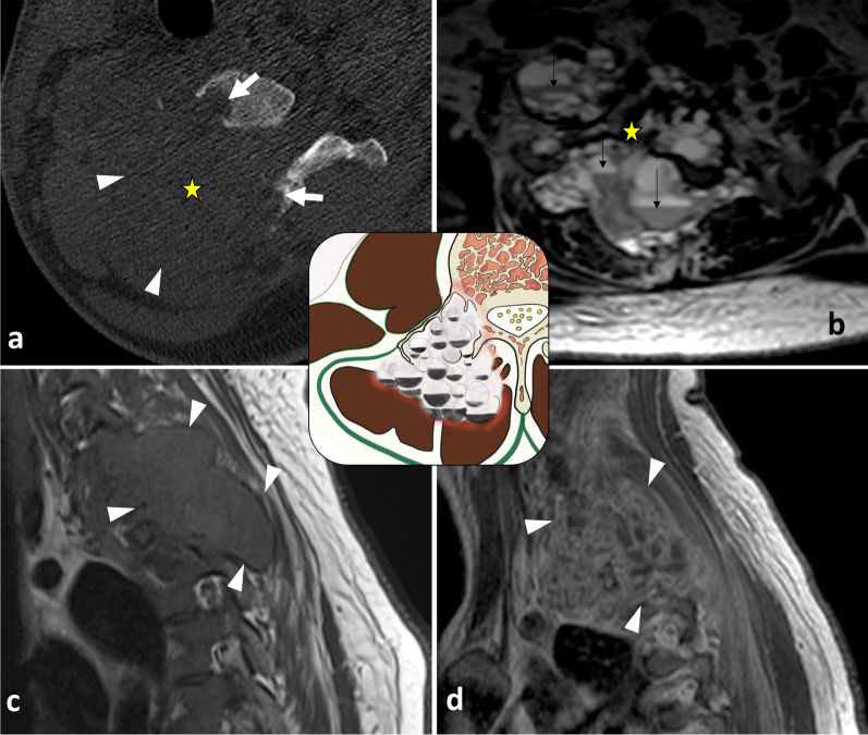
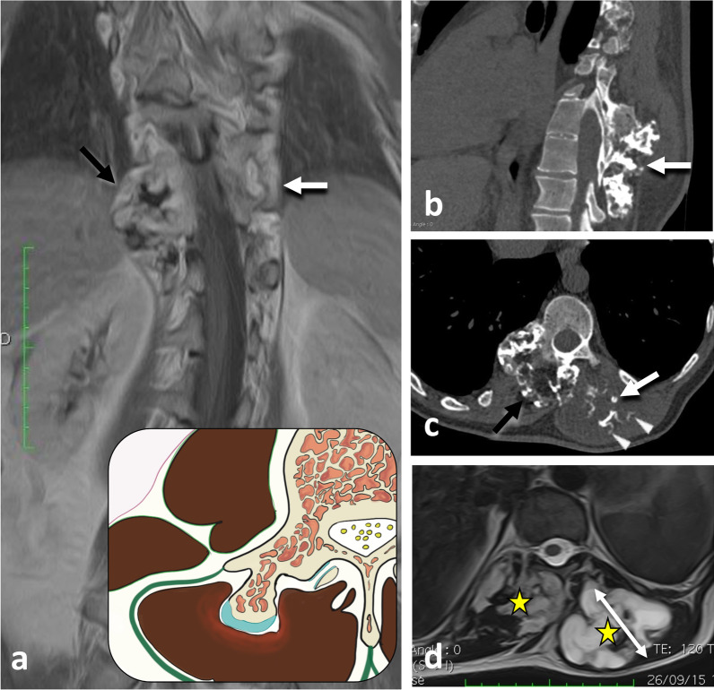
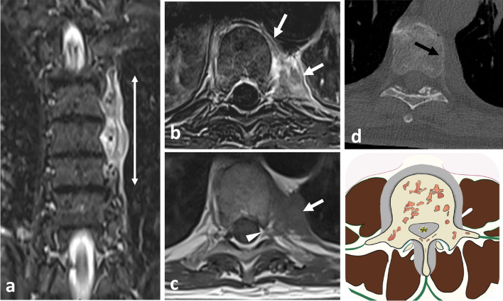
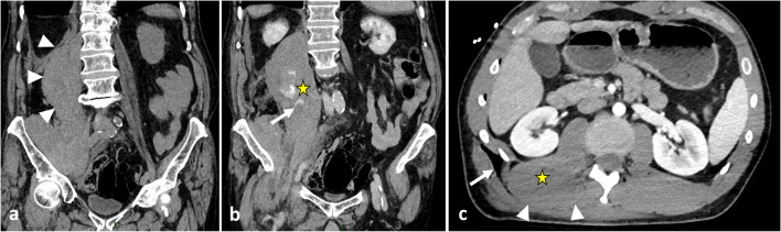
The paraspinal region encompasses all tissues around the spine. The regional anatomy is complex and includes the paraspinal muscles, spinal nerves, sympathetic chains, Batson's venous plexus and a rich arterial network. A wide variety of pathologies can occur in the paraspinal region, originating either from paraspinal soft tissues or the vertebral column. The most common paraspinal benign neoplasms include lipomas, fibroblastic tumours and benign peripheral nerve sheath tumours. Tumour-like masses such as haematomas, extramedullary haematopoiesis or abscesses should be considered in patients with suggestive medical histories. Malignant neoplasms are less frequent than benign processes and include liposarcomas and undifferentiated sarcomas. Secondary and primary spinal tumours may present as midline expansile soft tissue masses invading the adjacent paraspinal region. Knowledge of the anatomy of the paraspinal region is of major importance since it allows understanding of the complex locoregional tumour spread that can occur via many adipose corridors, haematogenous pathways and direct contact. Paraspinal tumours can extend into other anatomical regions, such as the retroperitoneum, pleura, posterior mediastinum, intercostal space or extradural neural axis compartment. Imaging plays a crucial role in formulating a hypothesis regarding the aetiology of the mass and tumour staging, which informs preoperative planning. Understanding the complex relationship between the different elements and the imaging features of common paraspinal masses is fundamental to achieving a correct diagnosis and adequate patient management. This review gives an overview of the anatomy of the paraspinal region and describes imaging features of the main tumours and tumour-like lesions that occur in the region.
椎旁区域包括脊柱周围的所有组织。该区域解剖结构复杂,包括椎旁肌、脊神经、交感神经链、巴特森静脉丛和丰富的动脉网络。椎旁区域可发生多种病理情况,起源于椎旁软组织或脊柱。最常见的椎旁良性肿瘤包括脂肪瘤、成纤维细胞肿瘤和良性周围神经鞘瘤。对于有提示性病史的患者,应考虑肿瘤样肿块,如血肿、髓外造血或脓肿。恶性肿瘤比良性病变少见,包括脂肪肉瘤和未分化肉瘤。继发性和原发性脊柱肿瘤可表现为中线膨胀性软组织肿块,侵犯相邻的椎旁区域。了解椎旁区域的解剖结构非常重要,因为它有助于理解肿瘤可能通过许多脂肪通道、血行途径和直接接触发生的复杂局部扩散。椎旁肿瘤可延伸至其他解剖区域,如腹膜后、胸膜、后纵隔、肋间间隙或硬脊膜外神经轴腔。影像学在形成关于肿块病因和肿瘤分期的假设方面起着关键作用,这为术前规划提供依据。了解不同结构之间的复杂关系以及常见椎旁肿块的影像学特征是实现正确诊断和妥善治疗患者的基础。本综述概述了椎旁区域的解剖结构,并描述了该区域主要肿瘤和肿瘤样病变的影像学特征。