School of Engineering, Center for Biomedical Engineering, Brown University, Providence, RI, USA.
Cardiovascular Research Center, Cardiovascular Institute, Rhode Island Hospital and Alpert Medical School of Brown University, Providence, RI, USA.
Methods Mol Biol. 2022;2485:147-157. doi: 10.1007/978-1-0716-2261-2_10.
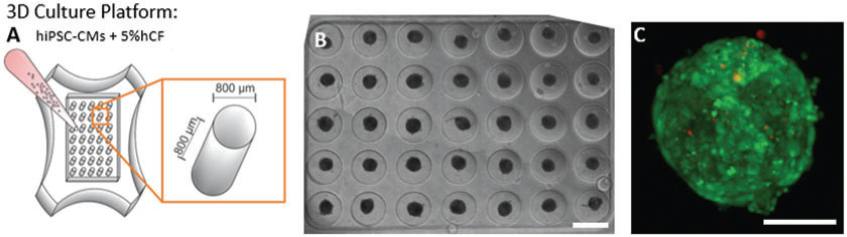
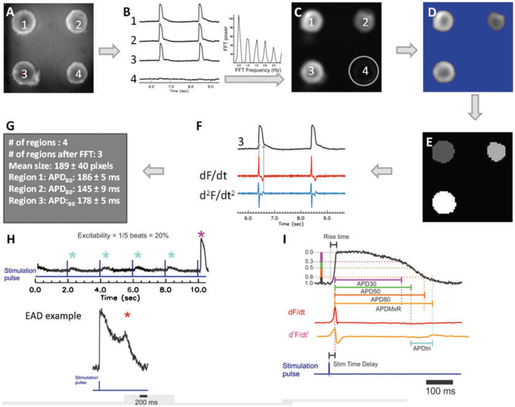
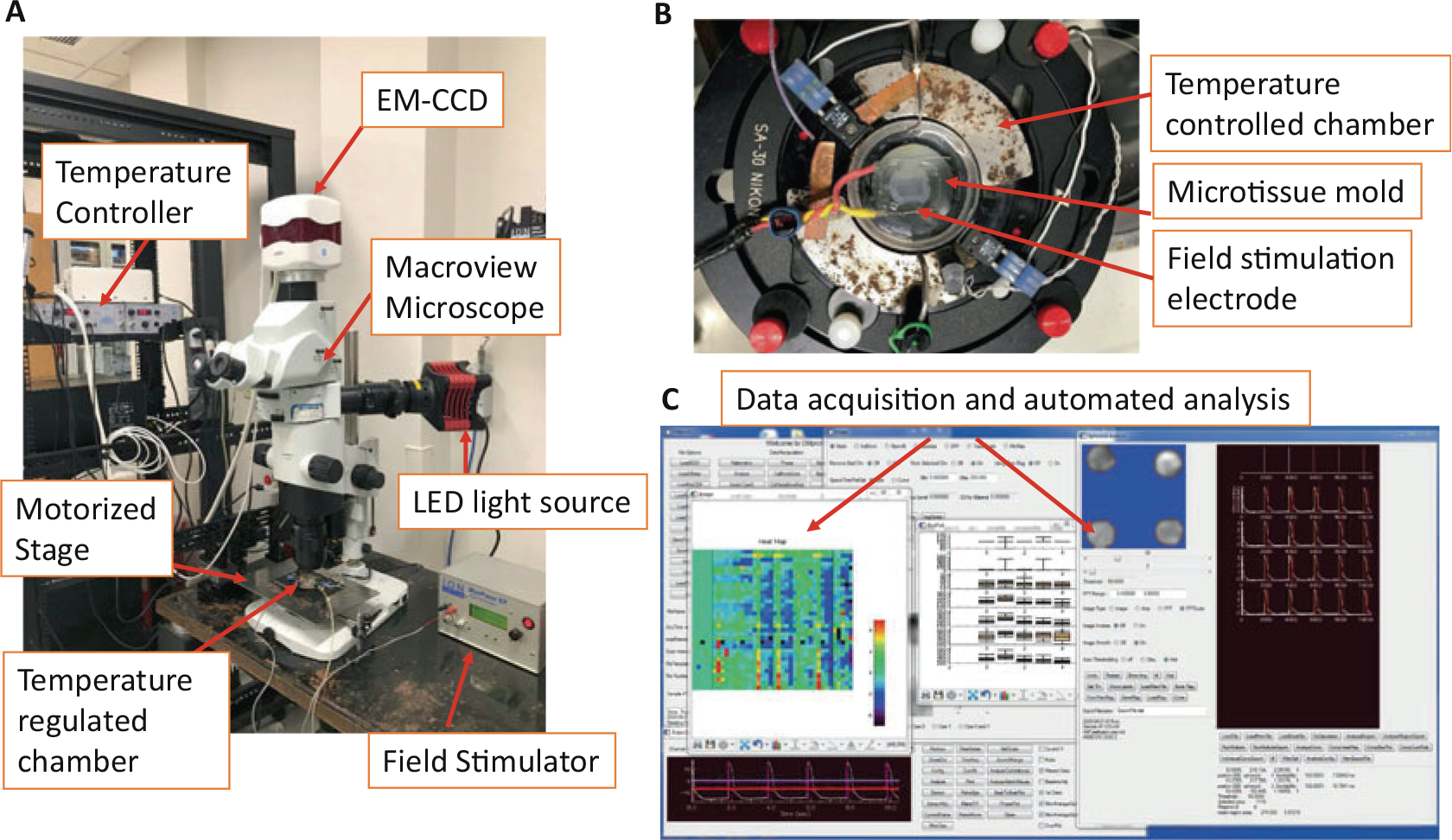
Risk assessment assays for chemically induced arrhythmia are critical, but significant limitations exist with current cardiotoxicity testing, including a focus on single select ion channels, the use of non-human species in vitro and in vivo, and limited direct physiological translation. To be predictive of actual adverse clinical arrhythmic risk, arrhythmia assessment models for chemicals and drugs should be fit-for-purpose and suited for evaluating compounds in which the mechanism of action may not be entirely known. Here, we describe methods for efficient and reliable screening for arrhythmogenic cardiotoxicity with a 3D human cardiac microtissue model using purified human-induced pluripotent stem cell (hiPSC)-derived cardiomyocytes and human cardiac fibroblasts. Applying optical mapping of voltage and calcium-sensitive dyes-an established approach to evaluate cardiac action potentials and calcium transients-to 3D heterotypic cardiac myocyte-fibroblast tissues allows for the generation and functional analysis of a large number of individual microtissues to provide greater throughput and high statistical power in analyses. Hundreds of microtissues in standard cell culture plates can be produced with low variability beat-to-beat, microtissue-to-microtissue, and across hiPSC-cardiomyocyte differentiation batches, reducing the number of microtissues required per condition for predictive outputs. The platform described here can be used as a sensitive, efficient, and predictive preclinical model validated for the purpose of assessing human pro-arrhythmic risk.
化学诱导性心律失常风险评估检测至关重要,但目前的心脏毒性测试存在显著局限性,包括仅关注单一选择离子通道、在体外和体内使用非人类物种,以及直接生理转化的有限性。为了预测实际的临床心律失常风险,化学物质和药物的心律失常评估模型应该具有针对性,适合评估作用机制不完全明确的化合物。在这里,我们描述了使用纯化的人诱导多能干细胞(hiPSC)衍生的心肌细胞和人心房成纤维细胞,通过 3D 人心肌微组织模型进行心律失常性心脏毒性的高效可靠筛选的方法。应用光学电压和钙敏染料标测——评估心脏动作电位和钙瞬变的既定方法——到 3 种异型性心肌细胞-成纤维细胞组织中,允许对大量单个微组织进行生成和功能分析,从而在分析中提供更高的通量和更高的统计能力。可以在标准细胞培养板中生成数百个具有低变异性的微组织,逐个微组织之间以及 hiPSC 心肌细胞分化批次之间的变异性都很小,从而减少了每个条件下用于预测输出所需的微组织数量。这里描述的平台可用作一种敏感、高效和可预测的临床前模型,经过验证可用于评估人类致心律失常风险。