Institute of Physiology, Faculty of Medicine, University of Duisburg-Essen, Hufelandstraße 55, D45147 Essen, Germany.
Institute of Medical Radiation Biology, Faculty of Medicine, University Duisburg-Essen, Hufelandstraße 55, D45147 Essen, Germany.
Cells. 2022 May 18;11(10):1671. doi: 10.3390/cells11101671.
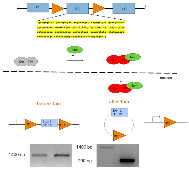
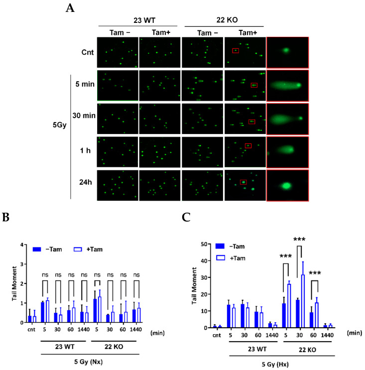
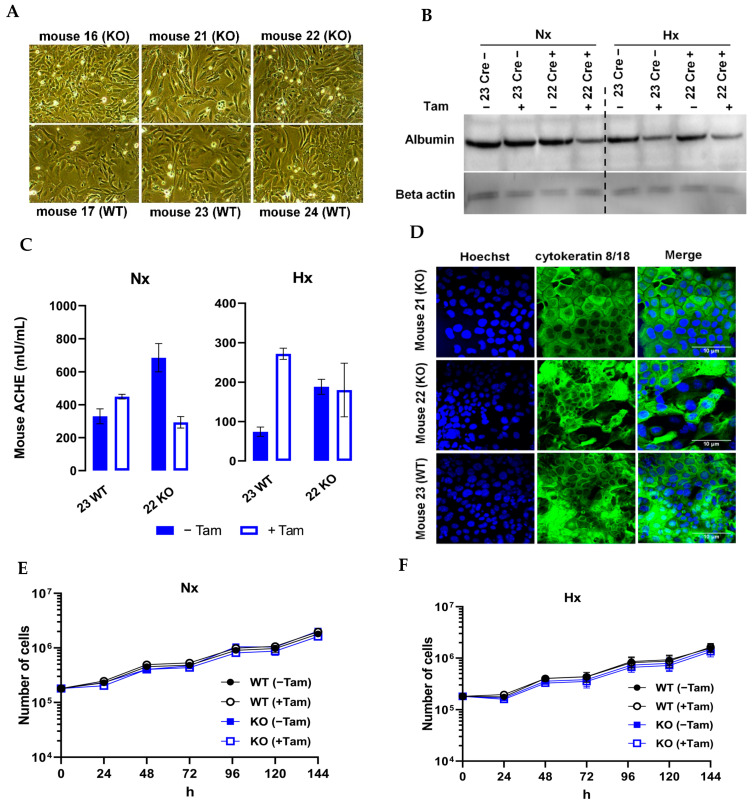
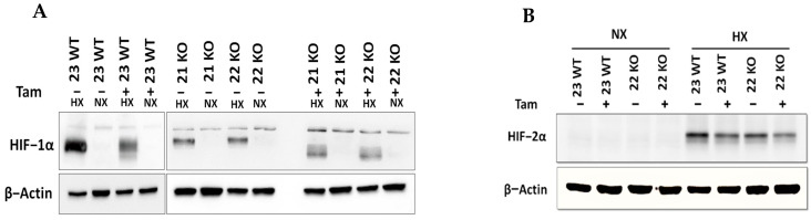
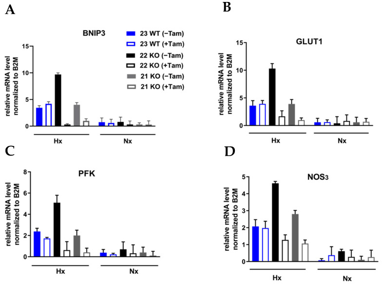
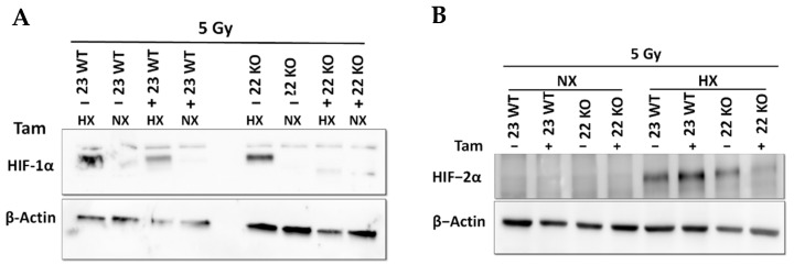
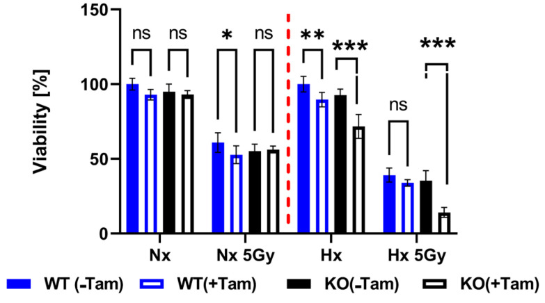
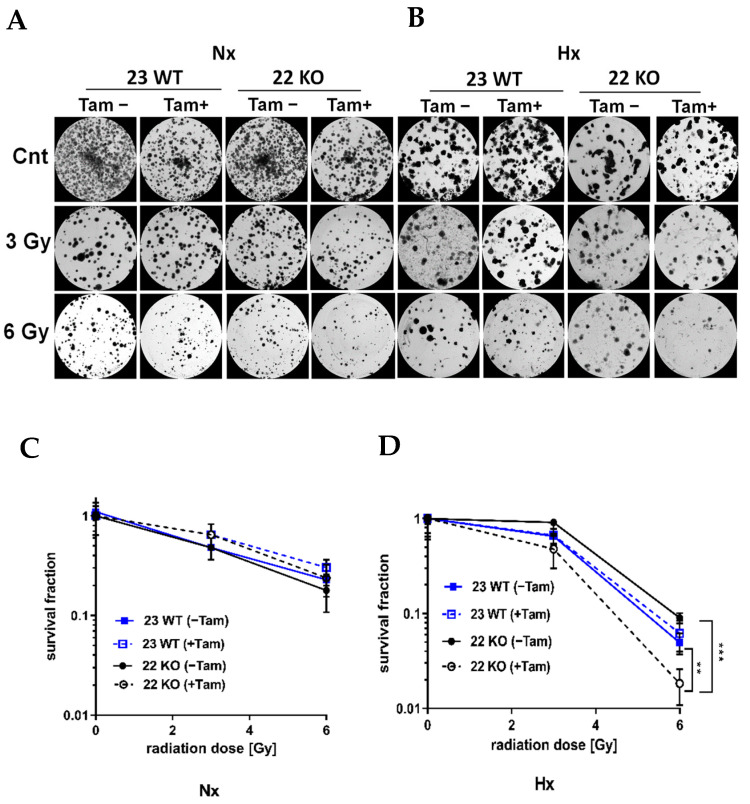
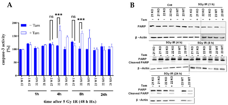
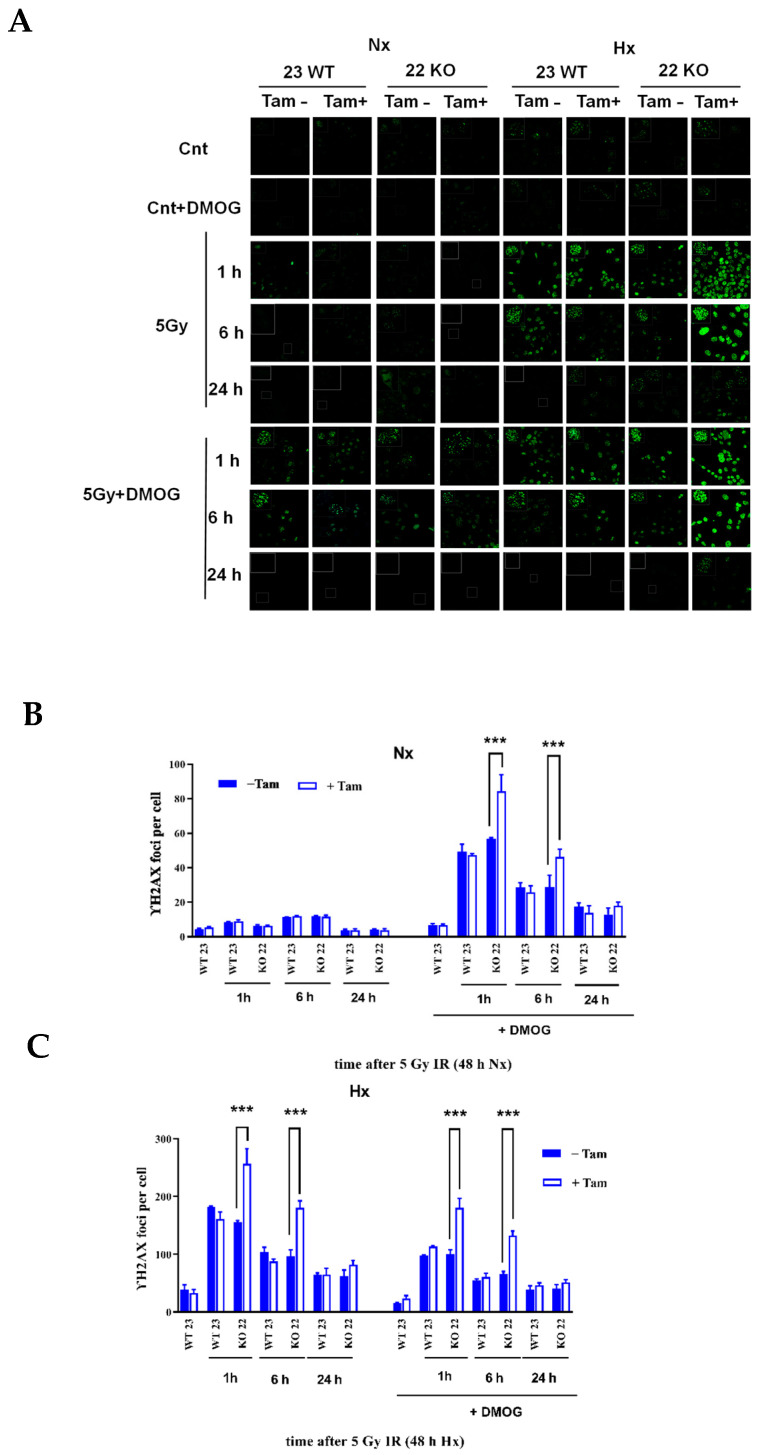
The transcription factor hypoxia-inducible factor (HIF) is the main oxygen sensor which regulates adaptation to cellular hypoxia. The aim of this study was to establish cultured murine hepatocyte derived cells (mHDC) as an in vitro model and to analyze the role of HIF-1α in apoptosis induction, DNA damage repair and sensitivity to ionizing radiation (IR). We have crossed C57/BL6 mice that bear loxP sites flanking exon 2 of Hif1a with mice which carry tamoxifen-inducible global Cre expression. From the offspring, we have established transduced hepatocyte cultures which are permanently HIF-1α deficient after tamoxifen treatment. We demonstrated that the cells produce albumin, acetylcholine esterase, and the cytokeratins 8 and 18 which functionally characterizes them as hepatocytes. In moderate hypoxia, HIF-1α deficiency increased IR-induced apoptosis and significantly reduced the surviving fraction of mHDC as compared to HIF-1α expressing cells in colony formation assays. Furthermore, HIF-1α knockout cells displayed increased IR-induced DNA damage as demonstrated by increased generation and persistence of γH2AX foci. HIF-1α deficient cells showed delayed DNA repair after IR in hypoxia in neutral comet assays which may indicate that non-homologous end joining (NHEJ) repair capacity was affected. Overall, our data suggest that HIF-1α inactivation increases radiation sensitivity of mHDC cells.
转录因子缺氧诱导因子 (HIF) 是调节细胞对缺氧适应的主要氧传感器。本研究的目的是建立培养的鼠源性肝细胞 (mHDC) 作为体外模型,并分析 HIF-1α 在细胞凋亡诱导、DNA 损伤修复和对电离辐射 (IR) 敏感性中的作用。我们将带有 Hif1a 外显子 2 侧翼loxP 位点的 C57/BL6 小鼠与携带他莫昔芬诱导的全局 Cre 表达的小鼠进行杂交。从后代中,我们建立了转导的肝细胞培养物,在他莫昔芬处理后永久性缺乏 HIF-1α。我们证明这些细胞产生白蛋白、乙酰胆碱酯酶和细胞角蛋白 8 和 18,这些功能特征使其成为肝细胞。在中度缺氧条件下,与表达 HIF-1α 的细胞相比,HIF-1α 缺失增加了 IR 诱导的细胞凋亡,并显著降低了 mHDC 的存活分数,在集落形成实验中。此外,HIF-1α 敲除细胞显示出更多的 IR 诱导的 DNA 损伤,如 γH2AX 焦点的产生和持续时间增加所示。在中性彗星试验中,缺氧条件下 HIF-1α 缺陷细胞在 IR 后 DNA 修复延迟,这可能表明非同源末端连接 (NHEJ) 修复能力受到影响。总的来说,我们的数据表明,HIF-1α 失活增加了 mHDC 细胞对辐射的敏感性。