Unit of Cardiology, Department of Traslational Medical Sciences, University of Campania "Luigi Vanvitelli", Monaldi Hospital, 80131 Naples, Italy.
Unit of Cardiology and Intensive Coronary Care, "Umberto I" Hospital, 84014 Nocera Inferiore, Italy.
Int J Environ Res Public Health. 2022 May 12;19(10):5898. doi: 10.3390/ijerph19105898.
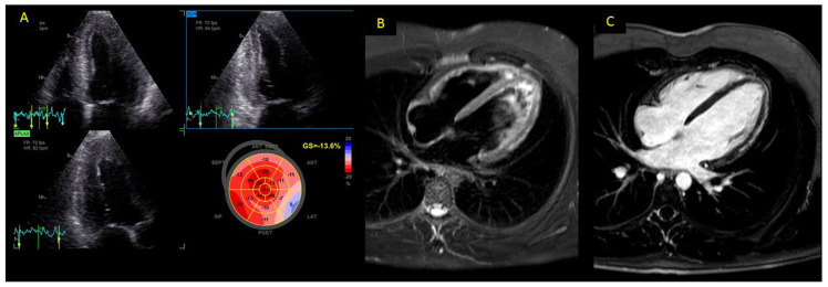
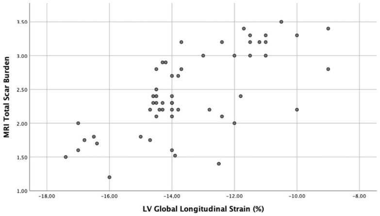
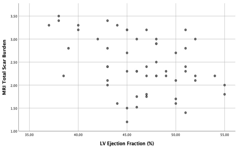
SARS-CoV2 infection, responsible for the COVID-19 disease, can determine cardiac as well as respiratory injury. In COVID patients, viral myocarditis can represent an important cause of myocardial damage. Clinical presentation of myocarditis is heterogeneous. Furthermore, the full diagnostic algorithm can be hindered by logistical difficulties related to the transportation of COVID-19 patients in a critical condition to the radiology department. Our aim was to study longitudinal systolic cardiac function in patients with COVID-19-related myocarditis with echocardiography and to compare these findings with cardiac magnetic resonance (CMR) results. Patients with confirmed acute myocarditis and age- and gender-matched healthy controls were enrolled. Both patients with COVID-19-related myocarditis and healthy controls underwent standard transthoracic echocardiography and speckle-tracking analysis at the moment of admission and after 6 months of follow-up. The data of 55 patients with myocarditis (mean age 46.4 ± 15.3, 70% males) and 55 healthy subjects were analyzed. The myocarditis group showed a significantly reduced global longitudinal strain (GLS) and sub-epicardial strain, compared to the control (p < 0.001). We found a positive correlation (r = 0.65, p < 0.0001) between total scar burden (TSB) on CMR and LV GLS. After 6 months of follow-up, GLS showed marked improvements in myocarditis patients on optimal medical therapy (p < 0.01). Furthermore, we showed a strong association between baseline GLS, left ventricular ejection fraction (LVEF) and TSB with LVEF at 6 months of follow-up. After a multivariable linear regression analysis, baseline GLS, LVEF and TSB were independent predictors of a functional outcome at follow-up (p < 0.0001). Cardiac function and myocardial longitudinal deformation, assessed by echocardiography, are associated with TSB at CMR and have a predictive value of functional recovery in the follow-up.
SARS-CoV2 感染是 COVID-19 疾病的病因,可导致心脏和呼吸系统损伤。在 COVID 患者中,病毒性心肌炎可能是心肌损伤的一个重要原因。心肌炎的临床表现具有异质性。此外,由于 COVID-19 患者在危急情况下向放射科转运的后勤困难,完整的诊断算法可能会受到阻碍。我们的目的是使用超声心动图研究 COVID-19 相关心肌炎患者的纵向收缩期心脏功能,并将这些发现与心脏磁共振(CMR)结果进行比较。纳入了确诊为急性心肌炎的患者和年龄、性别匹配的健康对照者。COVID-19 相关心肌炎患者和健康对照者均在入院时和 6 个月随访时接受标准经胸超声心动图和斑点追踪分析。对 55 例心肌炎患者(平均年龄 46.4 ± 15.3 岁,70%为男性)和 55 例健康受试者的数据进行了分析。与对照组相比,心肌炎组的整体纵向应变(GLS)和心外膜下应变明显降低(p < 0.001)。我们发现 CMR 上总瘢痕负荷(TSB)与 LV GLS 之间存在正相关(r = 0.65,p < 0.0001)。在接受最佳药物治疗后,心肌炎患者的 GLS 在 6 个月随访时显示出显著改善(p < 0.01)。此外,我们还发现基线 GLS、左心室射血分数(LVEF)和 TSB 与 6 个月随访时的 LVEF 之间存在很强的相关性。经过多变量线性回归分析,基线 GLS、LVEF 和 TSB 是随访时功能结果的独立预测因素(p < 0.0001)。超声心动图评估的心脏功能和心肌纵向变形与 CMR 上的 TSB 相关,并对随访时的功能恢复具有预测价值。