Pannone Luigi, Bisignani Antonio, Sorgente Antonio, Gauthey Anaïs, Della Rocca Domenico G, Monaco Cinzia, Bories Wim, Ramak Robbert, Overeinder Ingrid, Bala Gezim, Almorad Alexandre, Iacopino Saverio, Paparella Gaetano, Ströker Erwin, Sieira Juan, Flamée Panagiotis, Brugada Pedro, La Meir Mark, Chierchia Gian-Battista, De Asmundis Carlo
Heart Rhythm Management Centre, Postgraduate Program in Cardiac Electrophysiology and Pacing, Universitair Ziekenhuis Brussel, Vrije Universiteit Brussel, European Reference Networks Guard-Heart, Laarbeeklaan 101, 1090 Brussels, Belgium.
Anaesthesiology Department, Universitair Ziekenhuis Brussel, Vrije Universiteit Brussel, 1090 Brussels, Belgium.
J Clin Med. 2022 May 23;11(10):2948. doi: 10.3390/jcm11102948.
Non-contact charge density (CD) mapping allows a global visualization of left atrium (LA) activation and of activation patterns during atrial fibrillation (AF). The aim of this study was to analyze, with CD mapping, the changes in persistent AF induced by pulmonary vein isolation (PVI) and LA posterior wall isolation (LAPWI).
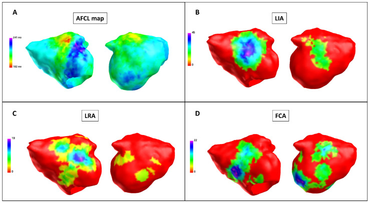
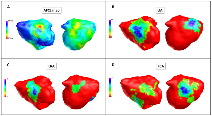
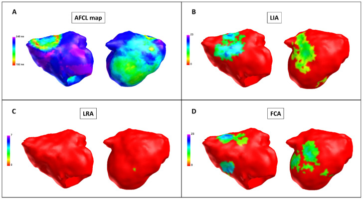
Patients undergoing PVI + LAPWI using the Arctic Front Advance PRO cryoballoon system were included in the study. CD maps were created during AF at baseline, after PVI and after LAPWI. Three distinct activation patterns were identified in the CD maps: localized irregular activation (LIA), localized rotational activation (LRA) and focal centrifugal activation (FCA). LA maps were divided into the following regions: anterior, septal, lateral, roof, posterior, inferior.
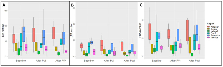
Eleven patients were included, with a total of 33 maps and 198 AF regions analyzed. Global and regional AF cycle lengths significantly increased after PVI and LAPWI. Baseline analysis demonstrated higher LIA, LRA and FCA numbers in the posterior and anterior regions. After PVI, there was no change in LIA, LRA and FCA occurrence. After PVI + LAPWI, a significant decrease in LRA was observed with no difference in LIA and FCA occurrence. In the regional analysis, there was a significant reduction in the LIA number in the inferior region, in the LRA number in the roof and posterior regions and in the FCA number in the lateral region.
A global reduction in the LRA number was observed only after PVI + LAPWI; it was driven by a reduction in rotational activity in the roof and posterior regions.
非接触式电荷密度(CD)标测可对左心房(LA)激活以及心房颤动(AF)期间的激活模式进行整体可视化。本研究旨在通过CD标测分析肺静脉隔离(PVI)和左心房后壁隔离(LAPWI)诱导的持续性房颤的变化。
纳入使用北极星前进PRO冷冻球囊系统进行PVI + LAPWI的患者。在房颤基线期、PVI后和LAPWI后创建CD图。在CD图中识别出三种不同的激活模式:局部不规则激活(LIA)、局部旋转激活(LRA)和局灶性离心激活(FCA)。左心房图分为以下区域:前部、间隔部、外侧、顶部、后部、下部。
纳入11例患者,共分析33张图和198个房颤区域。PVI和LAPWI后,整体和区域房颤周期长度显著增加。基线分析显示,后部和前部区域的LIA、LRA和FCA数量较多。PVI后,LIA、LRA和FCA的发生率没有变化。PVI + LAPWI后,观察到LRA显著减少,LIA和FCA的发生率没有差异。在区域分析中,下部区域的LIA数量、顶部和后部区域的LRA数量以及外侧区域的FCA数量显著减少。
仅在PVI + LAPWI后观察到LRA数量整体减少;这是由顶部和后部区域的旋转活动减少所致。