Carlsen Esben Andreas, Lindholm Kristian, Hindsholm Amalie, Gæde Mathias, Ladefoged Claes Nøhr, Loft Mathias, Johnbeck Camilla Bardram, Langer Seppo Wang, Oturai Peter, Knigge Ulrich, Kjaer Andreas, Andersen Flemming Littrup
Department of Clinical Physiology and Nuclear Medicine & Cluster for Molecular Imaging, Copenhagen University Hospital - Rigshospitalet & Department of Biomedical Sciences, University of Copenhagen, Copenhagen, Denmark.
ENETS Neuroendocrine Tumor Center of Excellence, Copenhagen University Hospital - Rigshospitalet, Copenhagen, Denmark.
EJNMMI Res. 2022 May 28;12(1):30. doi: 10.1186/s13550-022-00901-2.
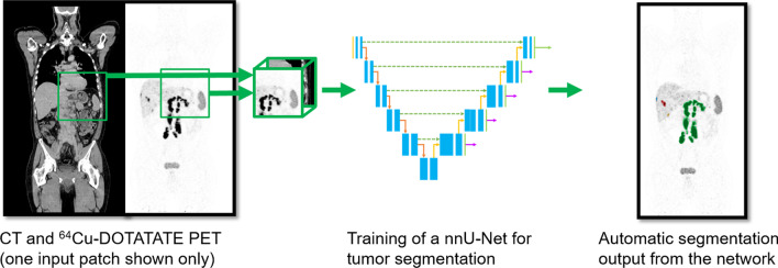
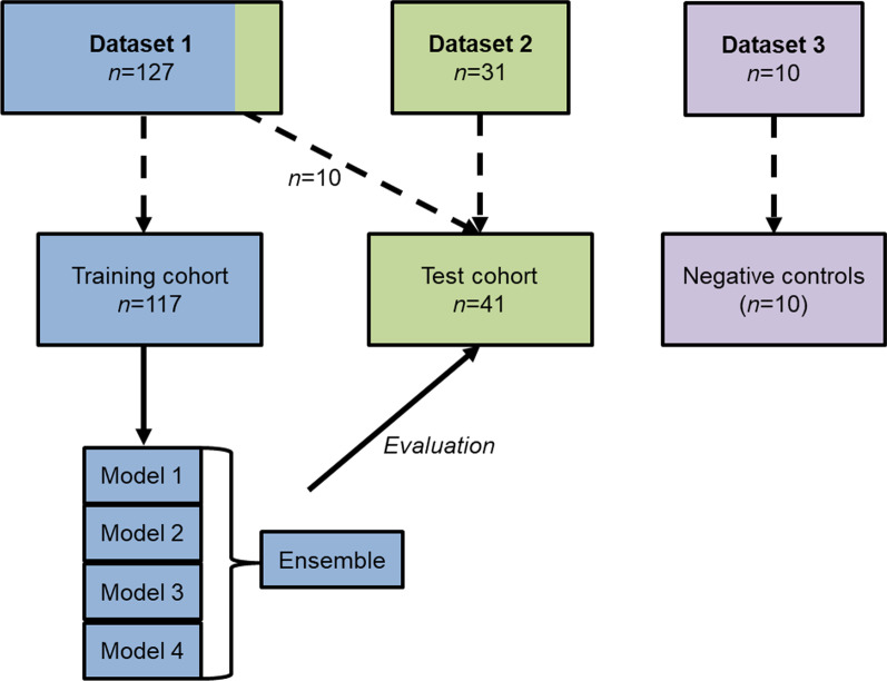
Segmentation of neuroendocrine neoplasms (NENs) in [Cu]Cu-DOTATATE positron emission tomography makes it possible to extract quantitative measures useable for prognostication of patients. However, manual tumor segmentation is cumbersome and time-consuming. Therefore, we aimed to implement and test an artificial intelligence (AI) network for tumor segmentation. Patients with gastroenteropancreatic or lung NEN with [Cu]Cu-DOTATATE PET/CT performed were included in our training (n = 117) and test cohort (n = 41). Further, 10 patients with no signs of NEN were included as negative controls. Ground truth segmentations were obtained by a standardized semiautomatic method for tumor segmentation by a physician. The nnU-Net framework was used to set up a deep learning U-net architecture. Dice score, sensitivity and precision were used for selection of the final model. AI segmentations were implemented in a clinical imaging viewer where a physician evaluated performance and performed manual adjustments.
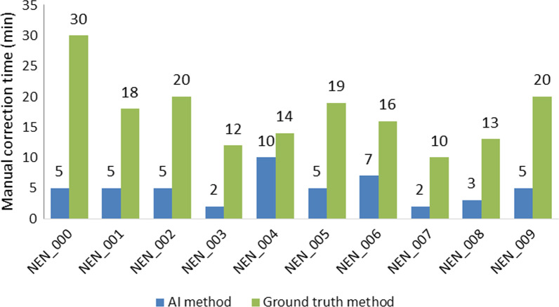
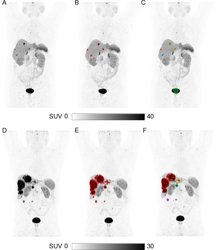
Cross-validation training was used to generate models and an ensemble model. The ensemble model performed best overall with a lesion-wise dice of 0.850 and pixel-wise dice, precision and sensitivity of 0.801, 0.786 and 0.872, respectively. Performance of the ensemble model was acceptable with some degree of manual adjustment in 35/41 (85%) patients. Final tumor segmentation could be obtained from the AI model with manual adjustments in 5 min versus 17 min for ground truth method, p < 0.01.
We implemented and validated an AI model that achieved a high similarity with ground truth segmentation and resulted in faster tumor segmentation. With AI, total tumor segmentation may become feasible in the clinical routine.
在[铜]铜-多柔比星正电子发射断层扫描中对神经内分泌肿瘤(NENs)进行分割,使得提取可用于患者预后评估的定量指标成为可能。然而,手动肿瘤分割既繁琐又耗时。因此,我们旨在实现并测试一种用于肿瘤分割的人工智能(AI)网络。纳入了接受过[铜]铜-多柔比星PET/CT检查的胃肠胰或肺NEN患者进入我们的训练队列(n = 117)和测试队列(n = 41)。此外,纳入10例无NEN迹象的患者作为阴性对照。通过医生采用标准化半自动方法进行肿瘤分割来获得真实分割结果。使用nnU-Net框架建立深度学习U-net架构。使用Dice分数、灵敏度和精确度来选择最终模型。AI分割在临床影像查看器中实现,医生在其中评估性能并进行手动调整。
采用交叉验证训练来生成模型和一个集成模型。集成模型总体表现最佳,病变层面的Dice值为0.850,像素层面的Dice值、精确度和灵敏度分别为0.801、0.786和0.872。在35/41(85%)的患者中,经过一定程度的手动调整后,集成模型的性能是可接受的。与真实分割方法相比,通过手动调整,AI模型在5分钟内即可获得最终肿瘤分割结果,而真实分割方法需要17分钟,p < 0.01。
我们实现并验证了一个与真实分割高度相似且能更快完成肿瘤分割的AI模型。借助AI,在临床常规工作中进行全肿瘤分割可能会变得可行。