• 文献检索
  • 文档翻译
  • 深度研究
  • 学术资讯
  • Suppr Zotero 插件Zotero 插件
  • 邀请有礼
  • 套餐&价格
  • 历史记录
应用&插件
Suppr Zotero 插件Zotero 插件浏览器插件Mac 客户端Windows 客户端微信小程序
定价
高级版会员购买积分包购买API积分包
服务
文献检索文档翻译深度研究API 文档MCP 服务
关于我们
关于 Suppr公司介绍联系我们用户协议隐私条款
关注我们

Suppr 超能文献

核心技术专利:CN118964589B侵权必究
粤ICP备2023148730 号-1Suppr @ 2026

文献检索

告别复杂PubMed语法,用中文像聊天一样搜索,搜遍4000万医学文献。AI智能推荐,让科研检索更轻松。

立即免费搜索

文件翻译

保留排版,准确专业,支持PDF/Word/PPT等文件格式,支持 12+语言互译。

免费翻译文档

深度研究

AI帮你快速写综述,25分钟生成高质量综述,智能提取关键信息,辅助科研写作。

立即免费体验

卷积神经网络在 PET 功能容积全自动分割中的应用:多中心环境下的开发和验证。

Convolutional neural networks for PET functional volume fully automatic segmentation: development and validation in a multi-center setting.

机构信息

LaTIM, INSERM, UMR 1101, University Brest, Brest, France.

GIGA-CRC in vivo Imaging, University of Liège, Liège, Belgium.

出版信息

Eur J Nucl Med Mol Imaging. 2021 Oct;48(11):3444-3456. doi: 10.1007/s00259-021-05244-z. Epub 2021 Mar 27.

DOI:10.1007/s00259-021-05244-z
PMID:33772335
原文链接:https://pmc.ncbi.nlm.nih.gov/articles/PMC8440243/
Abstract

PURPOSE

In this work, we addressed fully automatic determination of tumor functional uptake from positron emission tomography (PET) images without relying on other image modalities or additional prior constraints, in the context of multicenter images with heterogeneous characteristics.

METHODS

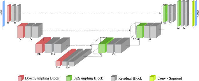
In cervical cancer, an additional challenge is the location of the tumor uptake near or even stuck to the bladder. PET datasets of 232 patients from five institutions were exploited. To avoid unreliable manual delineations, the ground truth was generated with a semi-automated approach: a volume containing the tumor and excluding the bladder was first manually determined, then a well-validated, semi-automated approach relying on the Fuzzy locally Adaptive Bayesian (FLAB) algorithm was applied to generate the ground truth. Our model built on the U-Net architecture incorporates residual blocks with concurrent spatial squeeze and excitation modules, as well as learnable non-linear downsampling and upsampling blocks. Experiments relied on cross-validation (four institutions for training and validation, and the fifth for testing).

RESULTS

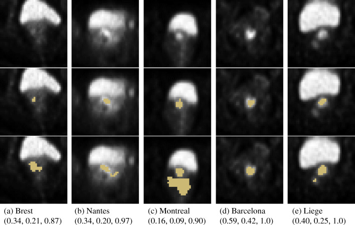
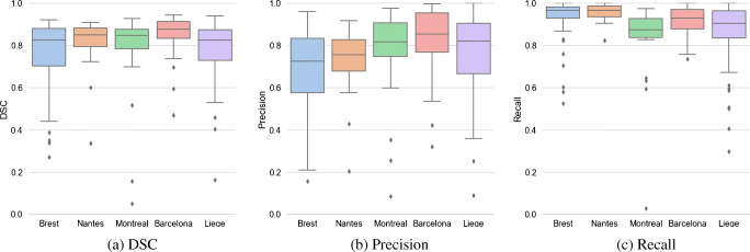
The model achieved good Dice similarity coefficient (DSC) with little variability across institutions (0.80 ± 0.03), with higher recall (0.90 ± 0.05) than precision (0.75 ± 0.05) and improved results over the standard U-Net (DSC 0.77 ± 0.05, recall 0.87 ± 0.02, precision 0.74 ± 0.08). Both vastly outperformed a fixed threshold at 40% of SUVmax (DSC 0.33 ± 0.15, recall 0.52 ± 0.17, precision 0.30 ± 0.16). In all cases, the model could determine the tumor uptake without including the bladder. Neither shape priors nor anatomical information was required to achieve efficient training.

CONCLUSION

The proposed method could facilitate the deployment of a fully automated radiomics pipeline in such a challenging multicenter context.

摘要

目的

在多中心具有异质特征的图像环境中,我们致力于在不依赖其他图像模态或额外先验约束的情况下,从正电子发射断层扫描(PET)图像中全自动确定肿瘤功能摄取。

方法

在宫颈癌中,肿瘤摄取的位置是一个额外的挑战,它靠近甚至紧贴膀胱。利用来自五个机构的 232 名患者的 PET 数据集。为了避免不可靠的手动勾画,使用半自动方法生成真实值:首先手动确定包含肿瘤且不包含膀胱的体积,然后应用经过良好验证的基于模糊局部自适应贝叶斯(FLAB)算法的半自动方法生成真实值。我们基于 U-Net 架构的模型包含带有并发空间挤压和激励模块的残差块,以及可学习的非线性下采样和上采样块。实验依赖于交叉验证(四个机构用于训练和验证,第五个机构用于测试)。

结果

该模型在不同机构之间具有较小的变异性,达到了良好的 Dice 相似系数(DSC)(0.80 ± 0.03),召回率(0.90 ± 0.05)高于精度(0.75 ± 0.05),并且优于标准 U-Net(DSC 0.77 ± 0.05,召回率 0.87 ± 0.02,精度 0.74 ± 0.08)。与 SUVmax 的 40%的固定阈值相比,两者的表现都要好得多(DSC 0.33 ± 0.15,召回率 0.52 ± 0.17,精度 0.30 ± 0.16)。在所有情况下,该模型都可以在不包含膀胱的情况下确定肿瘤摄取。既不需要形状先验,也不需要解剖信息来实现有效的训练。

结论

该方法可以促进在这种具有挑战性的多中心环境中部署全自动放射组学管道。

https://cdn.ncbi.nlm.nih.gov/pmc/blobs/488a/8440243/eb6f5d4a0d0d/259_2021_5244_Fig4_HTML.jpg
https://cdn.ncbi.nlm.nih.gov/pmc/blobs/488a/8440243/4fddc79fcc89/259_2021_5244_Fig1_HTML.jpg
https://cdn.ncbi.nlm.nih.gov/pmc/blobs/488a/8440243/e1c46007ca1c/259_2021_5244_Fig2_HTML.jpg
https://cdn.ncbi.nlm.nih.gov/pmc/blobs/488a/8440243/d7e800738883/259_2021_5244_Fig3_HTML.jpg
https://cdn.ncbi.nlm.nih.gov/pmc/blobs/488a/8440243/eb6f5d4a0d0d/259_2021_5244_Fig4_HTML.jpg

相似文献

1
Convolutional neural networks for PET functional volume fully automatic segmentation: development and validation in a multi-center setting.卷积神经网络在 PET 功能容积全自动分割中的应用:多中心环境下的开发和验证。
Eur J Nucl Med Mol Imaging. 2021 Oct;48(11):3444-3456. doi: 10.1007/s00259-021-05244-z. Epub 2021 Mar 27.
2
Fully automatic multi-organ segmentation for head and neck cancer radiotherapy using shape representation model constrained fully convolutional neural networks.使用基于形状表示模型约束的全卷积神经网络进行头颈部癌症放疗的全自动多器官分割。
Med Phys. 2018 Oct;45(10):4558-4567. doi: 10.1002/mp.13147. Epub 2018 Sep 19.
3
A Novel Framework for Automated Segmentation and Labeling of Homogeneous Versus Heterogeneous Lung Tumors in [F]FDG-PET Imaging.[F]FDG-PET成像中用于自动分割和标记同质与异质肺肿瘤的新型框架
Mol Imaging Biol. 2017 Jun;19(3):456-468. doi: 10.1007/s11307-016-1015-0.
4
The first MICCAI challenge on PET tumor segmentation.第一届 MICCAI 关于正电子发射断层扫描(PET)肿瘤分割挑战赛。
Med Image Anal. 2018 Feb;44:177-195. doi: 10.1016/j.media.2017.12.007. Epub 2017 Dec 9.
5
Fully automatic segmentation of diffuse large B cell lymphoma lesions on 3D FDG-PET/CT for total metabolic tumour volume prediction using a convolutional neural network.使用卷积神经网络对 3D FDG-PET/CT 上弥漫性大 B 细胞淋巴瘤病变进行全自动分割,以预测总代谢肿瘤体积。
Eur J Nucl Med Mol Imaging. 2021 May;48(5):1362-1370. doi: 10.1007/s00259-020-05080-7. Epub 2020 Oct 24.
6
The impact of multicentric datasets for the automated tumor delineation in primary prostate cancer using convolutional neural networks on F-PSMA-1007 PET.多中心数据集对使用卷积神经网络在F-PSMA-1007 PET上进行原发性前列腺癌自动肿瘤勾画的影响。
Radiat Oncol. 2024 Aug 7;19(1):106. doi: 10.1186/s13014-024-02491-w.
7
Automatic lesion detection and segmentation of 18F-FET PET in gliomas: A full 3D U-Net convolutional neural network study.基于全 3D U-Net 卷积神经网络的脑胶质瘤 18F-FET PET 自动病灶检测与分割研究。
PLoS One. 2018 Apr 13;13(4):e0195798. doi: 10.1371/journal.pone.0195798. eCollection 2018.
8
Fully Automatic Segmentation of Acute Ischemic Lesions on Diffusion-Weighted Imaging Using Convolutional Neural Networks: Comparison with Conventional Algorithms.基于卷积神经网络的弥散加权成像上急性缺血性病灶全自动分割:与传统算法的比较。
Korean J Radiol. 2019 Aug;20(8):1275-1284. doi: 10.3348/kjr.2018.0615.
9
A physics-guided modular deep-learning based automated framework for tumor segmentation in PET.基于物理引导的模块化深度学习的自动框架,用于 PET 中的肿瘤分割。
Phys Med Biol. 2020 Dec 18;65(24):245032. doi: 10.1088/1361-6560/ab8535.
10
An investigation of the effect of fat suppression and dimensionality on the accuracy of breast MRI segmentation using U-nets.利用 U-Nets 研究脂肪抑制和维度对乳腺 MRI 分割准确性的影响。
Med Phys. 2019 Mar;46(3):1230-1244. doi: 10.1002/mp.13375. Epub 2019 Feb 4.

引用本文的文献

1
Automatic cervical tumors segmentation in PET/MRI by parallel encoder U-net.基于并行编码器U型网络的PET/MRI图像中子宫颈肿瘤自动分割
Radiat Oncol. 2025 Jun 5;20(1):95. doi: 10.1186/s13014-025-02664-1.
2
Automatic detection and delineation of pediatric gliomas on combined [F]FET PET and MRI.基于联合[F]FET正电子发射断层显像(PET)与磁共振成像(MRI)的儿童胶质瘤自动检测与勾画
Front Nucl Med. 2022 Aug 24;2:960820. doi: 10.3389/fnume.2022.960820. eCollection 2022.
3
Cervical cancer segmentation based on medical images: a literature review.基于医学图像的宫颈癌分割:文献综述

本文引用的文献

1
Deep Learning-based Image Segmentation on Multimodal Medical Imaging.基于深度学习的多模态医学影像图像分割
IEEE Trans Radiat Plasma Med Sci. 2019 Mar;3(2):162-169. doi: 10.1109/trpms.2018.2890359. Epub 2019 Jan 1.
2
nnU-Net: a self-configuring method for deep learning-based biomedical image segmentation.nnU-Net:一种基于深度学习的生物医学图像分割的自配置方法。
Nat Methods. 2021 Feb;18(2):203-211. doi: 10.1038/s41592-020-01008-z. Epub 2020 Dec 7.
3
Variational PET/CT Tumor Co-segmentation Integrated with PET Restoration.
Quant Imaging Med Surg. 2024 Jul 1;14(7):5176-5204. doi: 10.21037/qims-24-369. Epub 2024 Jun 11.
4
Semi-supervised learning towards automated segmentation of PET images with limited annotations: application to lymphoma patients.半监督学习在有限标注 PET 图像自动分割中的应用:在淋巴瘤患者中的应用。
Phys Eng Sci Med. 2024 Sep;47(3):833-849. doi: 10.1007/s13246-024-01408-x. Epub 2024 Mar 21.
5
Machine Learning CT-Based Automatic Nodal Segmentation and PET Semi-Quantification of Intraoperative Ga-PSMA-11 PET/CT Images in High-Risk Prostate Cancer: A Pilot Study.基于机器学习的高危前列腺癌术中镓-PSMA-11 PET/CT图像的自动淋巴结分割及PET半定量分析:一项初步研究
Diagnostics (Basel). 2023 Sep 21;13(18):3013. doi: 10.3390/diagnostics13183013.
6
Joint EANM/SNMMI guideline on radiomics in nuclear medicine : Jointly supported by the EANM Physics Committee and the SNMMI Physics, Instrumentation and Data Sciences Council.核医学影像组学 EANM/SNMMI 指南:由 EANM 物理委员会和 SNMMI 物理、仪器和数据科学委员会共同支持。
Eur J Nucl Med Mol Imaging. 2023 Jan;50(2):352-375. doi: 10.1007/s00259-022-06001-6. Epub 2022 Nov 3.
7
An EANM position paper on the application of artificial intelligence in nuclear medicine.核医学中人工智能应用的 EANM 立场文件。
Eur J Nucl Med Mol Imaging. 2022 Dec;50(1):61-66. doi: 10.1007/s00259-022-05947-x. Epub 2022 Aug 25.
8
A convolutional neural network for total tumor segmentation in [Cu]Cu-DOTATATE PET/CT of patients with neuroendocrine neoplasms.用于神经内分泌肿瘤患者[铜]Cu-DOTATATE PET/CT中肿瘤全部分割的卷积神经网络。
EJNMMI Res. 2022 May 28;12(1):30. doi: 10.1186/s13550-022-00901-2.
9
Deep Learning With Radiomics for Disease Diagnosis and Treatment: Challenges and Potential.基于影像组学的深度学习在疾病诊断与治疗中的挑战与潜力
Front Oncol. 2022 Feb 17;12:773840. doi: 10.3389/fonc.2022.773840. eCollection 2022.
10
Accurate Tumor Delineation Rough Volume of Interest Analysis for F-FDG PET/CT Radiomics-Based Prognostic Modeling inNon-Small Cell Lung Cancer.非小细胞肺癌中基于F-FDG PET/CT影像组学的预后模型的精确肿瘤勾画及粗略感兴趣体积分析
Front Oncol. 2021 Oct 18;11:726865. doi: 10.3389/fonc.2021.726865. eCollection 2021.
结合PET图像重建的变分PET/CT肿瘤联合分割
IEEE Trans Radiat Plasma Med Sci. 2020 Jan;4(1):37-49. doi: 10.1109/trpms.2019.2911597. Epub 2019 Apr 16.
4
PET Image Denoising Using a Deep Neural Network Through Fine Tuning.通过微调深度学习网络实现PET图像去噪
IEEE Trans Radiat Plasma Med Sci. 2019 Mar;3(2):153-161. doi: 10.1109/TRPMS.2018.2877644. Epub 2018 Oct 23.
5
A physics-guided modular deep-learning based automated framework for tumor segmentation in PET.基于物理引导的模块化深度学习的自动框架,用于 PET 中的肿瘤分割。
Phys Med Biol. 2020 Dec 18;65(24):245032. doi: 10.1088/1361-6560/ab8535.
6
Improving Diagnostic Accuracy in Low-Dose SPECT Myocardial Perfusion Imaging With Convolutional Denoising Networks.卷积降噪网络提高低剂量 SPECT 心肌灌注成像的诊断准确性。
IEEE Trans Med Imaging. 2020 Sep;39(9):2893-2903. doi: 10.1109/TMI.2020.2979940. Epub 2020 Mar 10.
7
The effects of volume of interest delineation on MRI-based radiomics analysis: evaluation with two disease groups.基于感兴趣容积勾画对 MRI 影像组学分析的影响:两种疾病组的评估。
Cancer Imaging. 2019 Dec 21;19(1):89. doi: 10.1186/s40644-019-0276-7.
8
Estimates of incidence and mortality of cervical cancer in 2018: a worldwide analysis.2018 年宫颈癌发病率和死亡率的估计:全球分析。
Lancet Glob Health. 2020 Feb;8(2):e191-e203. doi: 10.1016/S2214-109X(19)30482-6. Epub 2019 Dec 4.
9
3D FULLY CONVOLUTIONAL NETWORKS FOR CO-SEGMENTATION OF TUMORS ON PET-CT IMAGES.用于PET-CT图像上肿瘤联合分割的3D全卷积网络
Proc IEEE Int Symp Biomed Imaging. 2018 Apr;2018:228-231. doi: 10.1109/ISBI.2018.8363561. Epub 2018 May 24.
10
Gross tumor volume segmentation for head and neck cancer radiotherapy using deep dense multi-modality network.使用深度密集多模态网络对头颈部癌症放射治疗的大体肿瘤体积分割。
Phys Med Biol. 2019 Oct 16;64(20):205015. doi: 10.1088/1361-6560/ab440d.