Department of Biochemistry, Kidwai Memorial Institute of Oncology Dr.M.H.Marigowda Road Bangalore, India.
Department of Biochemistry, Bengaluru Central University, Bangalore, India.
Asian Pac J Cancer Prev. 2022 May 1;23(5):1699-1709. doi: 10.31557/APJCP.2022.23.5.1699.
The study was aimed at understanding the survival of metastatic ovarian cancer spheroids in the malignant ascites microenvironment.
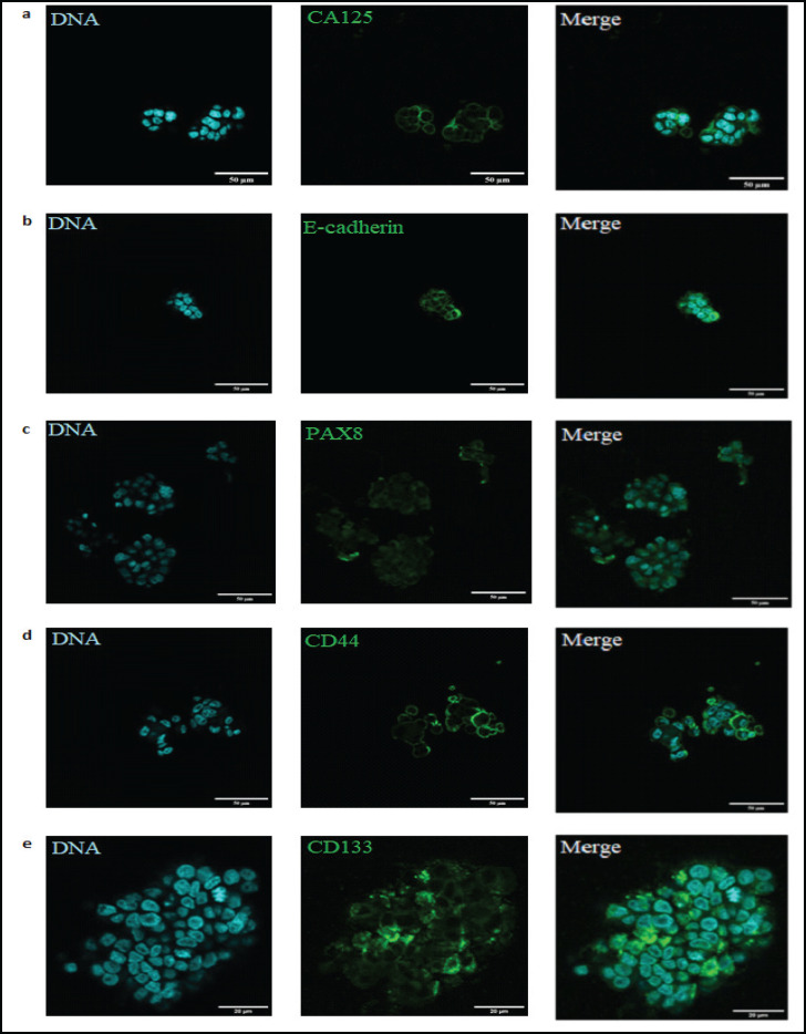
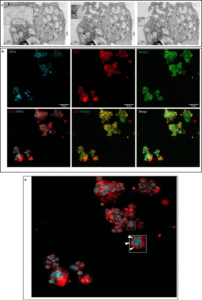
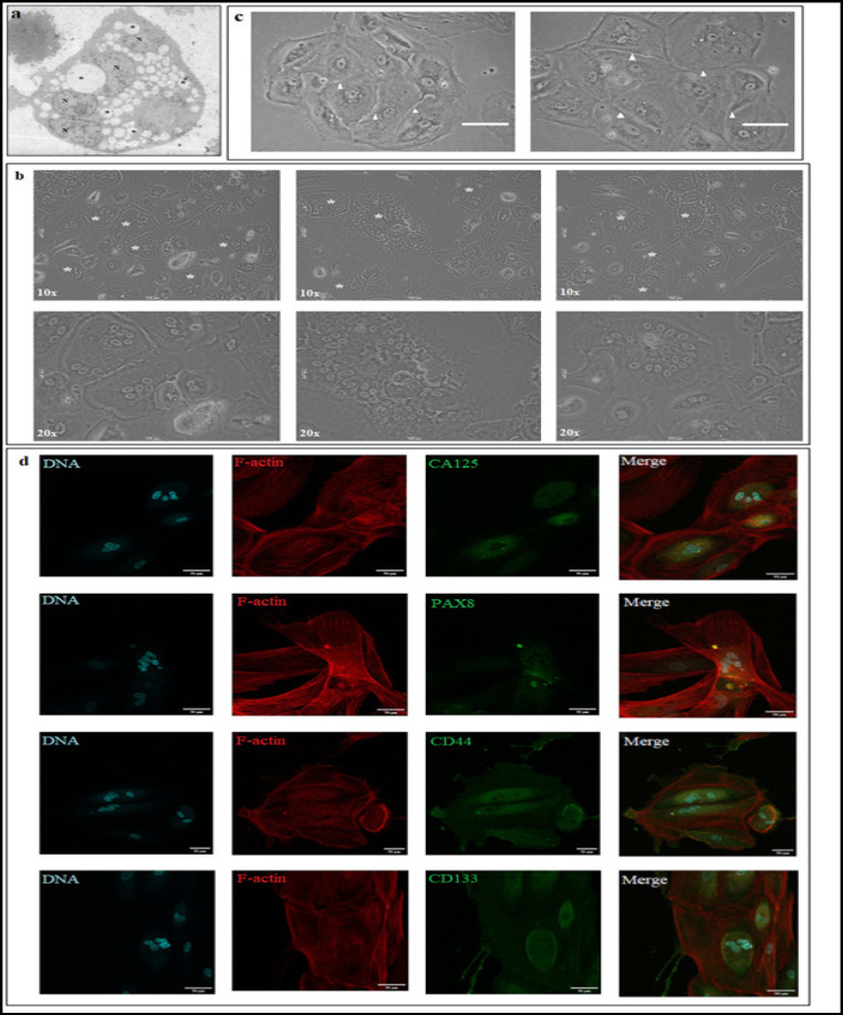
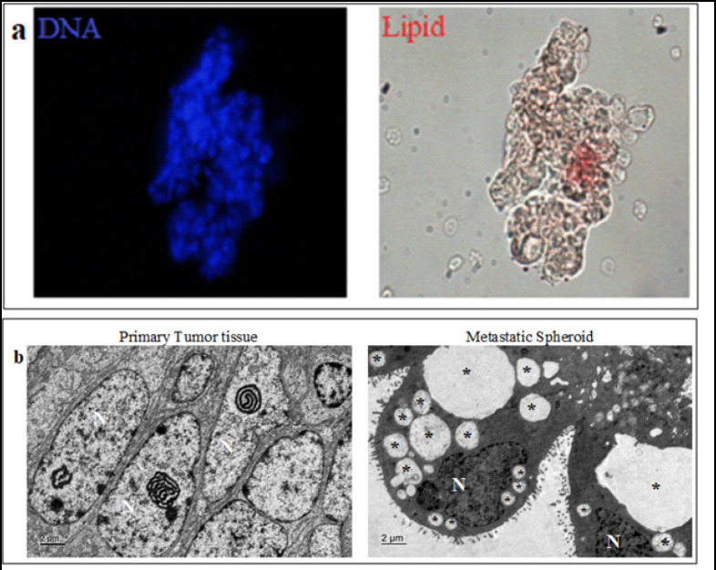
All the assays were performed using aseptically collected patient samples. The cells were characterized for the expression of ovarian and cancer stem cell markers using immunocytochemistry. The presence of lipid in the primary metastatic cancer spheroids were confirmed by neutral fat staining using Oil Red-O and transmission electron microscopy. The mRNA expression of autophagy and lipid metabolism genes was analyzed using RT-PCR. The lipid content was analyzed using lipidomics analysis. Etomoxir and chloroquine were used to study the effect of inhibition of autophagy in the metastatic cells. The data were analyzed using appropriate statistical tools and a p-value <0.05 was considered to be statistically significant.
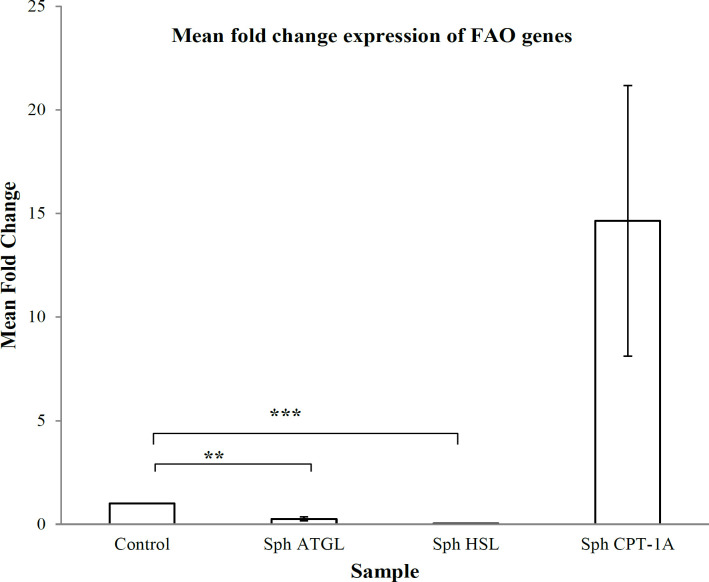
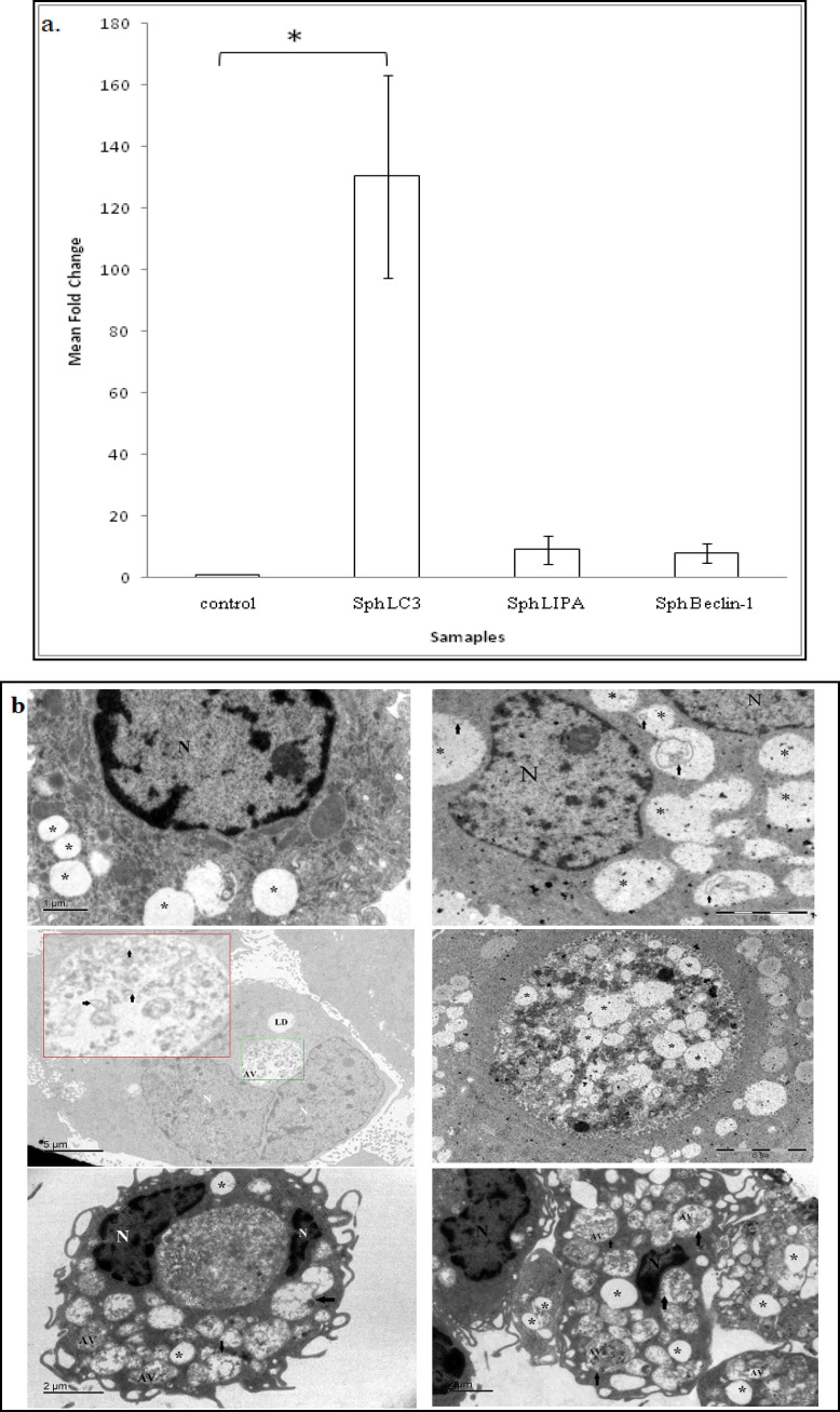
Metastatic ovarian cancer spheroids exhibit cancer stem like properties and undergo a metabolic reprogramming when they disseminate from the primary tumor. We report here the accumulation of numerous cytoplasmic lipid droplets and lipophagic vesicles in the metastatic cells in contrast to their primary tumors. In addition we also report that these cells depend on lipophagy for the utilization of lipids rather than the conventional lipolytic pathway. The lipidomics analysis data reveals that the metastatic cells possess high levels of unsaturated fatty acids. We have also reported the occurrence of distinct accumulation of multiple nuclei in the patient derived metastatic cells. Inhibition of beta-oxidation and autophagic machinery using etomoxir and chloroquine resulted in cell death suggesting a potential mode to suppress metastatic cancer cells.
Metabolic reprogramming is a characteristic feature of the metastatic ovarian cancer cells that are persisting in the malignant ascites. Targeting of the metastatic by gaining an insight into the various metabolic and molecular changes that occur in the metastatic niche provides a promising therapeutic approach in management of the disease.
本研究旨在了解转移性卵巢癌球体在恶性腹水微环境中的存活情况。
所有实验均使用无菌采集的患者样本进行。通过免疫细胞化学法检测卵巢和癌症干细胞标志物的表达来鉴定细胞。使用油红 O 中性脂肪染色和透射电子显微镜确认原发性转移性癌症球体中脂质的存在。使用 RT-PCR 分析自噬和脂质代谢基因的 mRNA 表达。使用脂质组学分析来分析脂质含量。使用乙莫克司和氯喹研究抑制转移性细胞自噬的效果。使用适当的统计工具进行数据分析,p 值<0.05 被认为具有统计学意义。
转移性卵巢癌球体在从原发性肿瘤扩散时表现出类似癌症干细胞的特性,并经历代谢重编程。我们在这里报告了与原发性肿瘤相比,转移性细胞中大量细胞质脂质滴和噬脂体的积累。此外,我们还报告说,这些细胞依赖于脂噬作用来利用脂质,而不是传统的脂肪分解途径。脂质组学分析数据显示,转移性细胞具有高水平的不饱和脂肪酸。我们还报告了在患者来源的转移性细胞中发生的多个独特核的明显积累。使用乙莫克司和氯喹抑制β-氧化和自噬机制会导致细胞死亡,这表明抑制转移性癌细胞的潜在模式。
代谢重编程是在恶性腹水中持续存在的转移性卵巢癌细胞的特征。通过深入了解转移性部位发生的各种代谢和分子变化来靶向转移性,为该疾病的管理提供了有前途的治疗方法。