National Center for Pleura and Peritoneum, NCT South-West Germany, Tübingen, Germany.
Department of Preclinical Imaging and Radiopharmacy, Werner Siemens Imaging Center, University Hospital Tübingen, Tübingen, Germany.
J Transl Med. 2022 Dec 12;20(1):581. doi: 10.1186/s12967-022-03763-3.
The poor prognosis of ovarian cancer patients is strongly related to peritoneal metastasis with the production of malignant ascites. However, it remains largely unclear how ascites in the peritoneal cavity influences tumor metabolism and recurrence. This study is an explorative approach aimed at for a deeper molecular and physical-chemical characterization of malignant ascites and to investigate their effect on in vitro ovarian cancer cell proliferation.
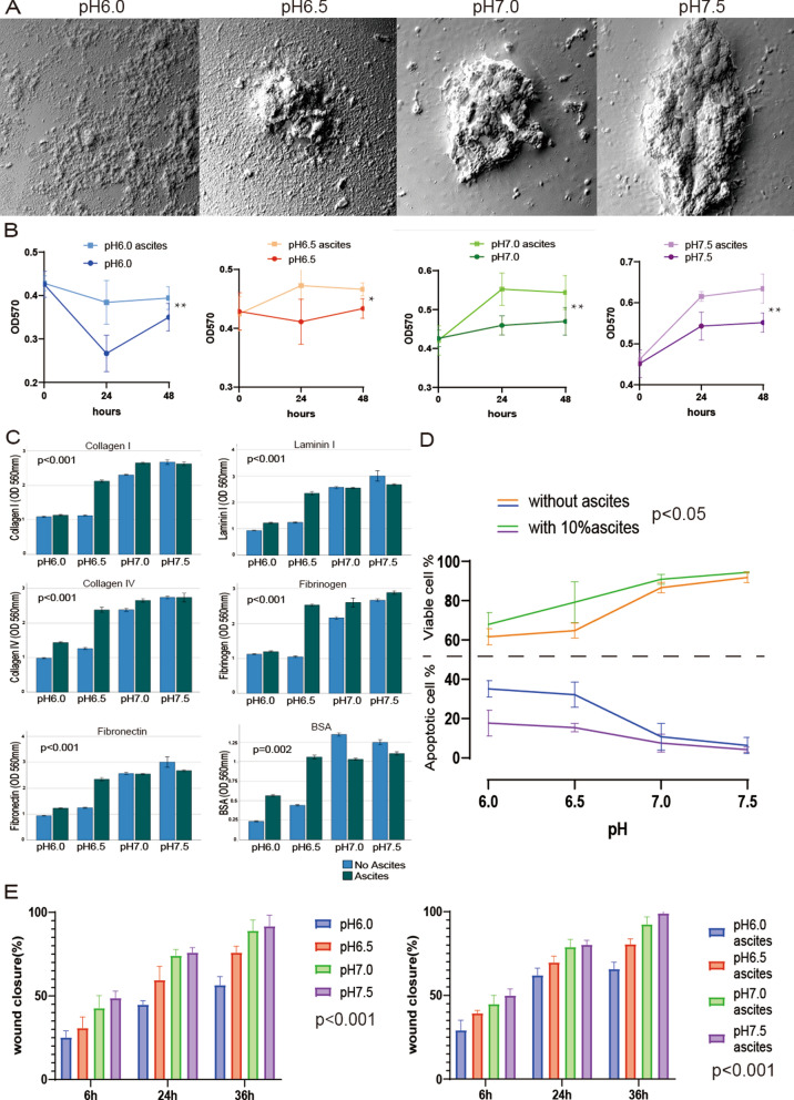
This study included 10 malignant ascites specimens from patients undergoing ovarian cancer resection. Ascites samples were deeply phenotyped by H-NMR based metabolomics, blood-gas analyzer based gas flow analysis and flow cytomertry based a 13-plex cytokine panel. Characteristics of tumor cells were investigated in a 3D spheroid model by SEM and metabolic activity, adhesion, anti-apoptosis, migratory ability evaluated by MTT assay, adhesion assay, flowcytometry and scratch assay. The effect of different pH values was assessed by adding 10% malignant ascites to the test samples.
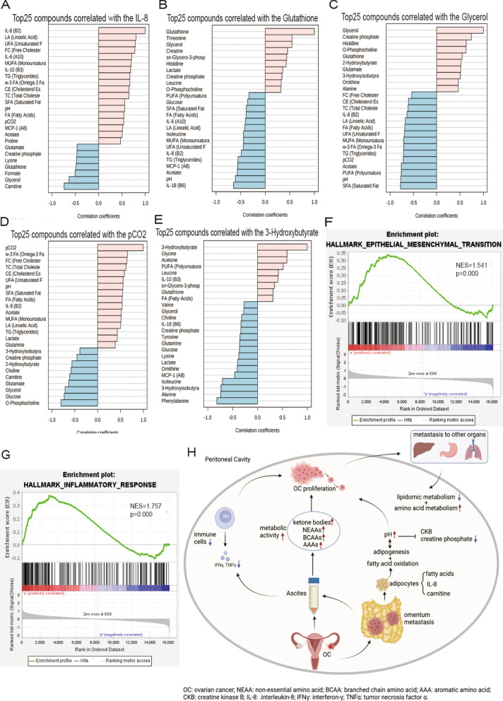
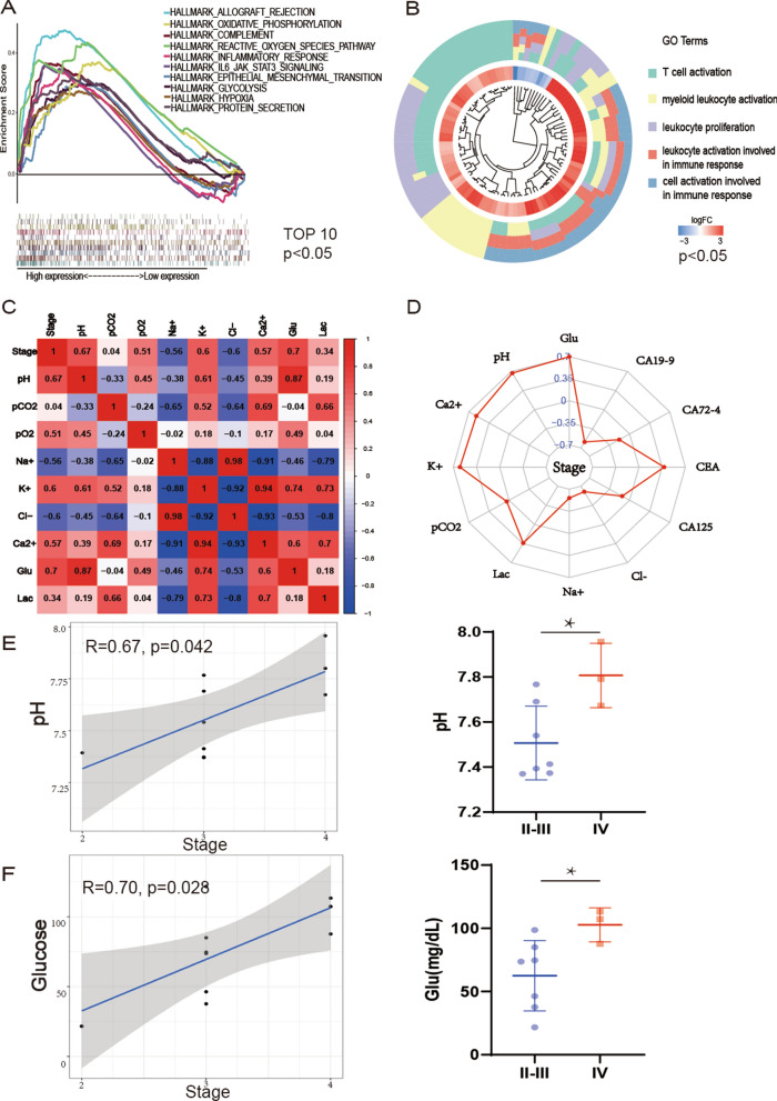
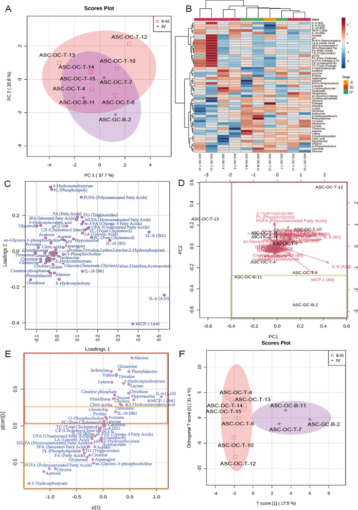
The overall extracellular (peritoneal) environment was alkaline, with pH of ascites at stage II-III = 7.51 ± 0.16, and stage IV = 7.78 ± 0.16. Ovarian cancer spheroids grew rapidly in a slightly alkaline environment. Decreasing pH of the cell culture medium suppressed tumor features, metabolic activity, adhesion, anti-apoptosis, and migratory ability. However, 10% ascites could prevent tumor cells from being affected by acidic pH. Metabolomics analysis identified stage IV patients had significantly higher concentrations of alanine, isoleucine, phenylalanine, and glutamine than stage II-III patients, while stage II-III patients had significantly higher concentrations of 3-hydroxybutyrate. pH was positively correlated with acetate, and acetate positively correlated with lipid compounds. IL-8 was positively correlated with lipid metabolites and acetate. Glutathione and carnitine were negatively correlated with cytokines IL-6 and chemokines (IL-8 & MCP-1).
Alkaline malignant ascites facilitated ovarian cancer progression. Additionally, deep ascites phenotyping by metabolomics and cytokine investigations allows for a refined stratification of ovarian cancer patients. These findings contribute to the understanding of ascites pathology in ovarian cancer.
卵巢癌患者预后不良与腹膜转移和恶性腹水的产生密切相关。然而,腹水如何影响肿瘤代谢和复发仍在很大程度上不清楚。本研究旨在更深入地研究恶性腹水的分子和物理化学特性,并研究其对体外卵巢癌细胞增殖的影响,是一种探索性方法。
本研究纳入了 10 例接受卵巢癌切除术的患者的恶性腹水标本。通过基于 H-NMR 的代谢组学、血气分析仪的气流分析和基于流式细胞术的 13 个细胞因子面板对腹水样本进行了深入的表型分析。通过 SEM 观察肿瘤细胞在 3D 球体模型中的特征,并通过 MTT 测定、粘附测定、流式细胞术和划痕试验评估代谢活性、粘附、抗凋亡和迁移能力。通过向测试样本中添加 10%恶性腹水来评估不同 pH 值的影响。
总的细胞外(腹膜)环境呈碱性,II-III 期腹水的 pH 值为 7.51±0.16,IV 期腹水的 pH 值为 7.78±0.16。卵巢癌球体在略碱性环境中生长迅速。降低细胞培养液的 pH 值抑制了肿瘤特征、代谢活性、粘附、抗凋亡和迁移能力。然而,10%的腹水可以防止肿瘤细胞受到酸性 pH 的影响。代谢组学分析表明,IV 期患者的丙氨酸、异亮氨酸、苯丙氨酸和谷氨酰胺浓度明显高于 II-III 期患者,而 II-III 期患者的 3-羟基丁酸浓度明显更高。pH 值与乙酸呈正相关,乙酸与脂质化合物呈正相关。IL-8 与脂质代谢物和乙酸呈正相关。谷胱甘肽和肉碱与细胞因子 IL-6 和趋化因子(IL-8 和 MCP-1)呈负相关。
碱性恶性腹水促进了卵巢癌的进展。此外,通过代谢组学和细胞因子研究对腹水进行深入表型分析,可以对卵巢癌患者进行更精细的分层。这些发现有助于了解卵巢癌腹水的病理。