Singh Priyanka, Saha Sonali, Tripathi Abhay Mani, Yadav Gunjan, Dhinsa Kavita
Department of Pedodontics and Preventive Dentistry, Sardar Patel Post Graduate Institute of Dental Sciences, Lucknow, Uttar Pradesh, India.
Int J Clin Pediatr Dent. 2022;15(Suppl 1):S22-S29. doi: 10.5005/jp-journals-10005-2126.
To evaluate root canal transportation, centering ability ratio (CAR), remaining dentine thickness, dentinal cracks, and instrumentation time after instrumentation with different filing systems in root canals of primary teeth by cone beam computed tomography (CBCT) analysis.
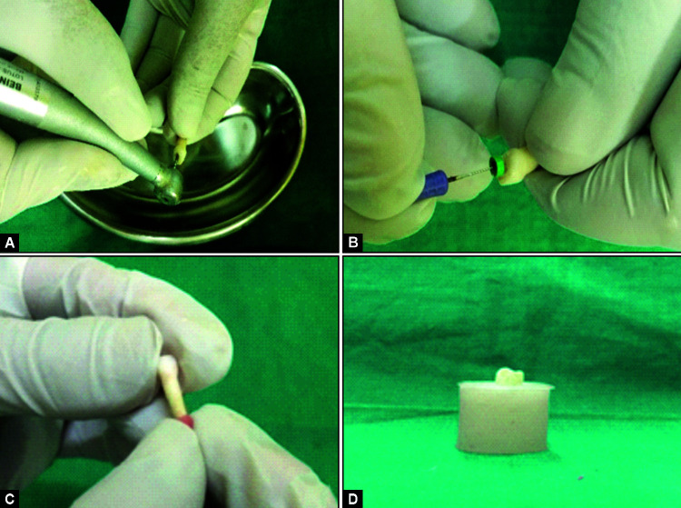
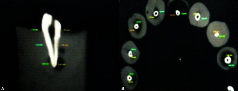
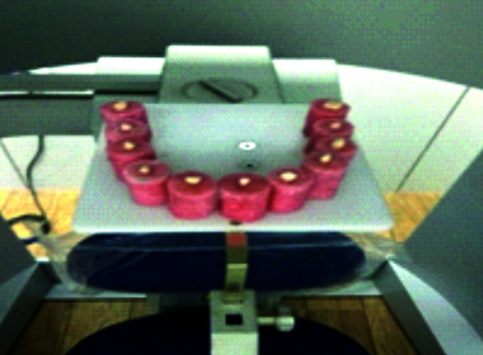
Sixty prepared canals of primary teeth divided into 4 groups with 15 canals in each were prepared with NiTi K files, Proaper Next (PTN) files, OneShape (OS), and WaveOne (WO) files, respectively. Using CBCT scan, the pre- and postinstrumentation scan was done to obtain images at three levels (apical, middle, and cervical). The results obtained were statistically analyzed using SPSS 21 statistical software version.
Significant statistical difference was found between different filing systems.
ProTaper Next files showed least canal transportation and the best centering ability was shown by OS file system. The NiTi K hand files preserved maximum remaining dentin thickness (RDT) and produced minimum dentin cracks. WO file system took least instrumentation time when compared to the other three filing systems.
The use of rotary instruments in the pulpectomy of primary teeth represents a promising technique being advantageous for the pediatric patients by maintaining the original canal curvatures, showing greater ability to maintain dentin thickness, causing lesser dentin cracks, and reducing chair time thus favoring preparation of more conical root canals and better obturation
Singh P. Cone-beam Computed Tomographic Analysis of Deciduous Root Canals after Instrumentation with Different Filing Systems: An Study. Int J Clin Pediatr Dent. 2022;15(S-1):S22-S29.
通过锥形束计算机断层扫描(CBCT)分析,评估不同锉系统对乳牙根管进行预备后根管偏移、定心能力比(CAR)、剩余牙本质厚度、牙本质裂纹及预备时间。
60颗已预备的乳牙根管分为4组,每组15个根管,分别用镍钛K锉、ProTaper Next(PTN)锉、OneShape(OS)锉和WaveOne(WO)锉进行预备。使用CBCT扫描,在预备前后进行扫描以获取根尖、中部和颈部三个层面的图像。使用SPSS 21统计软件版本对所得结果进行统计学分析。
不同锉系统之间存在显著统计学差异。
ProTaper Next锉显示出最小的根管偏移,OS锉系统表现出最佳的定心能力。镍钛K手用锉保留了最大的剩余牙本质厚度(RDT),并产生最少的牙本质裂纹。与其他三种锉系统相比,WO锉系统的预备时间最短。
在乳牙牙髓摘除术中使用旋转器械是一种有前景的技术,对儿童患者有利,因为它能保持原始根管弯曲度,具有更强的保持牙本质厚度的能力,产生更少的牙本质裂纹,并减少椅旁时间,从而有利于制备更呈锥形的根管和更好的根管充填。
Singh P. 不同锉系统预备后乳牙根管的锥形束计算机断层扫描分析:一项研究。《国际临床儿科牙科学杂志》。2022;15(S-1):S22-S29。