• 文献检索
  • 文档翻译
  • 深度研究
  • 学术资讯
  • Suppr Zotero 插件Zotero 插件
  • 邀请有礼
  • 套餐&价格
  • 历史记录
应用&插件
Suppr Zotero 插件Zotero 插件浏览器插件Mac 客户端Windows 客户端微信小程序
定价
高级版会员购买积分包购买API积分包
服务
文献检索文档翻译深度研究API 文档MCP 服务
关于我们
关于 Suppr公司介绍联系我们用户协议隐私条款
关注我们

Suppr 超能文献

核心技术专利:CN118964589B侵权必究
粤ICP备2023148730 号-1Suppr @ 2026

文献检索

告别复杂PubMed语法,用中文像聊天一样搜索,搜遍4000万医学文献。AI智能推荐,让科研检索更轻松。

立即免费搜索

文件翻译

保留排版,准确专业,支持PDF/Word/PPT等文件格式,支持 12+语言互译。

免费翻译文档

深度研究

AI帮你快速写综述,25分钟生成高质量综述,智能提取关键信息,辅助科研写作。

立即免费体验

术前评估肝细胞癌的组织学分级和微血管浸润的临床和影像学特征。

Clinical and imaging features preoperative evaluation of histological grade and microvascular infiltration of hepatocellular carcinoma.

机构信息

Department of Radiology, Nanfang Hospital, Southern Medical University, Guangzhou, 510515, China.

NanFang PET Center, Nanfang Hospital, Southern Medical University, 1838 Guangzhou Avenue North, Guangzhou, 510515, Guangdong, China.

出版信息

BMC Gastroenterol. 2022 Aug 1;22(1):369. doi: 10.1186/s12876-022-02449-w.

DOI:10.1186/s12876-022-02449-w
PMID:35915440
原文链接:https://pmc.ncbi.nlm.nih.gov/articles/PMC9341046/
Abstract

BACKGROUND

To predict the histological grade and microvascular invasion (MVI) in patients with HCC.

METHODS

A retrospective analysis was conducted on 175 patients who underwent MRI enhancement scanning (from September 2016.9 to October 2020). They were divided into MVI positive, MVI negative, Grade-high and Grade-low groups.

RESULTS

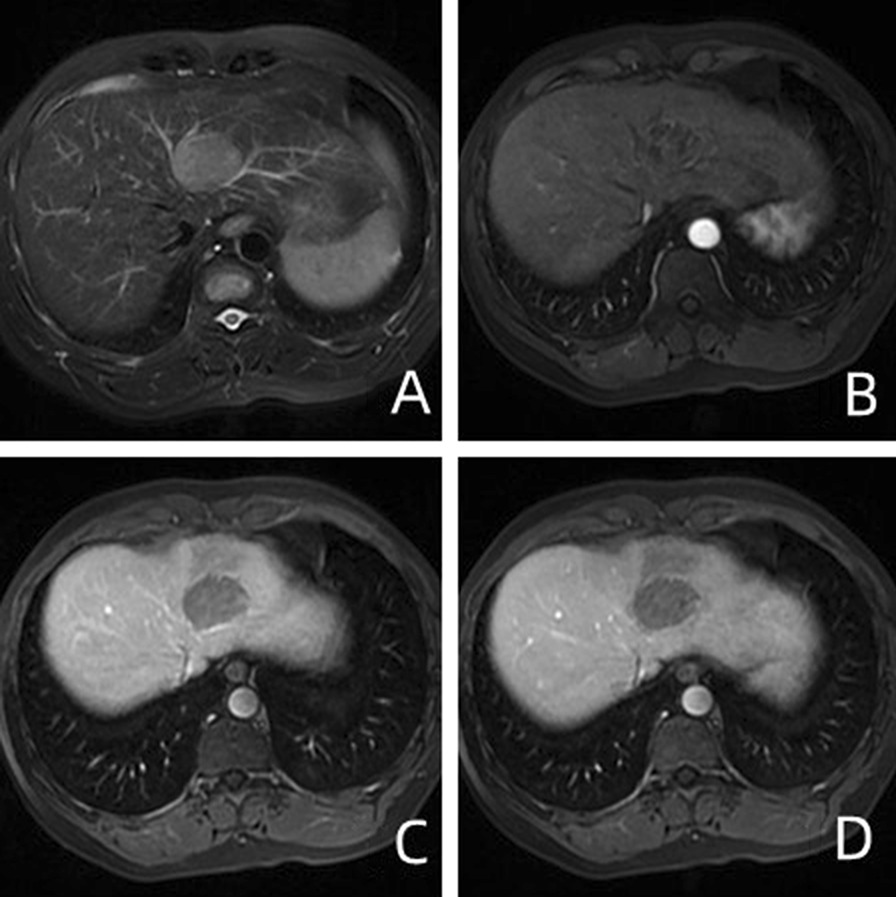
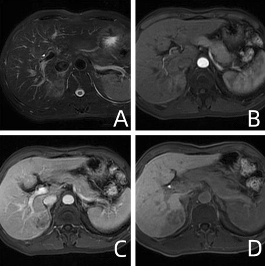
The AFP of 175 HCC patients distributed in MVI positive and negative groups, Grade-low and Grade-high groups were statistically significant (P = 0.002 and 0.03, respectively). Multiple HCC lesions were more common in MVI positive and Grade-high groups. Correspondingly, more single lesions were found in MVI negative and Grade-low groups (P = 0.005 and 0.019, respectively). Capsule on MRI was more common in MVI negative and Grade-high groups, and the difference was statistically significant (P = 0.02 and 0.011, respectively). There were statistical differences in the distribution of three MRI signs: artistic rim enhancement, artistic peripheral enhancement, and tumor margin between MVI positive and MVI negative groups (P = 0.001, < 0.001, and < 0.001, respectively). Tumor hypointensity on HBP was significantly different between MVI positive and negative groups (P < 0.001).

CONCLUSIONS

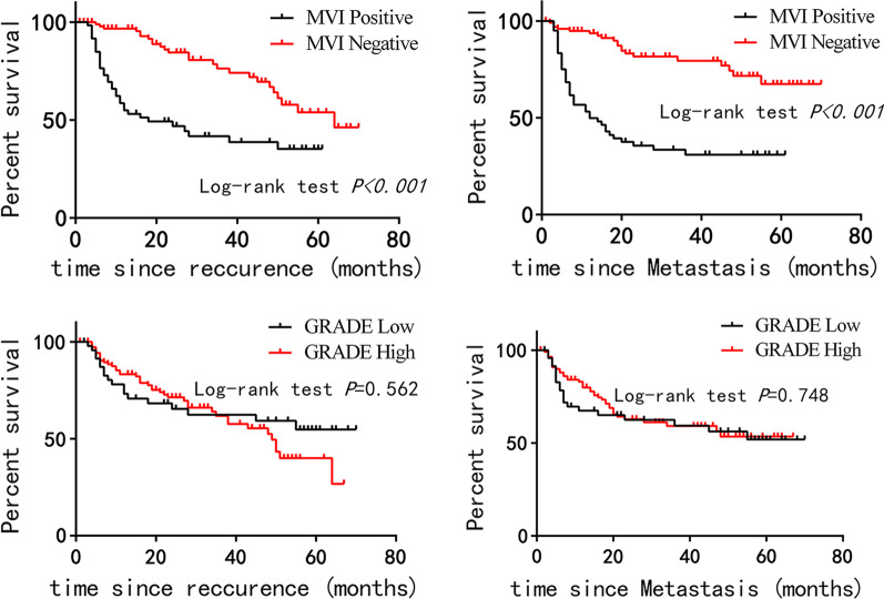
Our research shows that preoperative enhanced imaging can be used to predict MVI and tumor differentiation grade of HCC. The prognosis of MVI-negative group was better than that of MVI-positive group.

摘要

背景

预测 HCC 患者的组织学分级和微血管侵犯(MVI)。

方法

回顾性分析了 175 例接受 MRI 增强扫描的患者(2016 年 9 月至 2020 年 10 月)。他们分为 MVI 阳性、MVI 阴性、高分级和低分级组。

结果

175 例 HCC 患者的 AFP 在 MVI 阳性和阴性组、低分级和高分级组之间存在统计学差异(P=0.002 和 0.03)。MVI 阳性和高分级组中多发 HCC 病变更为常见,而 MVI 阴性和低分级组中则更为常见单发性病变(P=0.005 和 0.019)。MRI 上的包膜在 MVI 阴性和高分级组中更为常见,差异具有统计学意义(P=0.02 和 0.011)。MVI 阳性和阴性组之间存在三种 MRI 征象的分布差异:艺术边缘增强、艺术周边增强和肿瘤边缘(P=0.001、<0.001 和<0.001)。HBP 上肿瘤低信号在 MVI 阳性和阴性组之间有显著差异(P<0.001)。

结论

我们的研究表明,术前增强成像可用于预测 HCC 的 MVI 和肿瘤分化程度。MVI 阴性组的预后优于 MVI 阳性组。

https://cdn.ncbi.nlm.nih.gov/pmc/blobs/1a59/9341046/ed81ac6d486a/12876_2022_2449_Fig3_HTML.jpg
https://cdn.ncbi.nlm.nih.gov/pmc/blobs/1a59/9341046/3ed3b9afda07/12876_2022_2449_Fig1_HTML.jpg
https://cdn.ncbi.nlm.nih.gov/pmc/blobs/1a59/9341046/cd87f77ce2c3/12876_2022_2449_Fig2_HTML.jpg
https://cdn.ncbi.nlm.nih.gov/pmc/blobs/1a59/9341046/ed81ac6d486a/12876_2022_2449_Fig3_HTML.jpg
https://cdn.ncbi.nlm.nih.gov/pmc/blobs/1a59/9341046/3ed3b9afda07/12876_2022_2449_Fig1_HTML.jpg
https://cdn.ncbi.nlm.nih.gov/pmc/blobs/1a59/9341046/cd87f77ce2c3/12876_2022_2449_Fig2_HTML.jpg
https://cdn.ncbi.nlm.nih.gov/pmc/blobs/1a59/9341046/ed81ac6d486a/12876_2022_2449_Fig3_HTML.jpg

相似文献

1
Clinical and imaging features preoperative evaluation of histological grade and microvascular infiltration of hepatocellular carcinoma.术前评估肝细胞癌的组织学分级和微血管浸润的临床和影像学特征。
BMC Gastroenterol. 2022 Aug 1;22(1):369. doi: 10.1186/s12876-022-02449-w.
2
Hepatocellular carcinoma: preoperative gadoxetic acid-enhanced MR imaging can predict early recurrence after curative resection using image features and texture analysis.肝细胞癌:术前钆塞酸增强磁共振成像可通过图像特征和纹理分析预测根治性切除术后的早期复发。
Abdom Radiol (NY). 2019 Feb;44(2):539-548. doi: 10.1007/s00261-018-1768-9.
3
A model incorporating histopathology and preoperative gadoxetic acid-enhanced MRI to predict early recurrence of hepatocellular carcinoma without microvascular invasion after curative hepatectomy.纳入组织病理学和术前钆塞酸增强 MRI 的模型预测根治性肝切除术后无微血管侵犯的肝细胞癌早期复发。
Br J Radiol. 2023 Mar 1;96(1144):20220739. doi: 10.1259/bjr.20220739. Epub 2023 Mar 6.
4
Preoperative gadoxetic acid-enhanced MRI for predicting microvascular invasion in patients with single hepatocellular carcinoma.术前钆塞酸增强 MRI 预测单发肝细胞癌患者的微血管侵犯。
J Hepatol. 2017 Sep;67(3):526-534. doi: 10.1016/j.jhep.2017.04.024. Epub 2017 May 6.
5
Prediction of HCC microvascular invasion with gadobenate-enhanced MRI: correlation with pathology.钆贝葡胺增强 MRI 预测 HCC 微血管侵犯:与病理学的相关性。
Eur Radiol. 2020 Oct;30(10):5327-5336. doi: 10.1007/s00330-020-06895-6. Epub 2020 May 4.
6
The value of quantitative MR elastography-based stiffness for assessing the microvascular invasion grade in hepatocellular carcinoma.基于定量磁共振弹性成像的硬度在评估肝细胞癌微血管侵犯分级中的价值。
Eur Radiol. 2023 Jun;33(6):4103-4114. doi: 10.1007/s00330-022-09290-5. Epub 2022 Nov 26.
7
IVIM improves preoperative assessment of microvascular invasion in HCC.多体素 IVIM 模型有助于术前评估 HCC 的微血管侵犯。
Eur Radiol. 2019 Oct;29(10):5403-5414. doi: 10.1007/s00330-019-06088-w. Epub 2019 Mar 15.
8
Gadoxetic acid-enhanced MRI as a predictor of recurrence of HCC after liver transplantation.钆塞酸增强 MRI 作为肝移植后 HCC 复发的预测指标。
Eur Radiol. 2020 Feb;30(2):987-995. doi: 10.1007/s00330-019-06424-0. Epub 2019 Aug 30.
9
Radiomic analysis based on multi-phase magnetic resonance imaging to predict preoperatively microvascular invasion in hepatocellular carcinoma.基于多期磁共振成像的放射组学分析预测肝细胞癌术前微血管侵犯。
World J Gastroenterol. 2022 Jun 28;28(24):2733-2747. doi: 10.3748/wjg.v28.i24.2733.
10
ADC similarity predicts microvascular invasion of bifocal hepatocellular carcinoma.ADC 相似性预测双焦点肝细胞癌的微血管侵犯。
Abdom Radiol (NY). 2018 Sep;43(9):2295-2302. doi: 10.1007/s00261-018-1469-4.

引用本文的文献

1
Differentiating Liver Metastases from Primary Liver Cancer: A Retrospective Study of Imaging and Pathological Features in Patients with Histopathological Confirmation.鉴别肝转移瘤与原发性肝癌:一项对经组织病理学确诊患者的影像学和病理特征的回顾性研究
Biomedicines. 2025 Jan 11;13(1):164. doi: 10.3390/biomedicines13010164.
2
Cross-institutional evaluation of deep learning and radiomics models in predicting microvascular invasion in hepatocellular carcinoma: validity, robustness, and ultrasound modality efficacy comparison.跨机构评估深度学习和放射组学模型在预测肝细胞癌微血管侵犯中的应用:有效性、稳健性和超声模态效能比较。
Cancer Imaging. 2024 Oct 22;24(1):142. doi: 10.1186/s40644-024-00790-9.
3

本文引用的文献

1
The Roles of Diffusion Kurtosis Imaging and Intravoxel Incoherent Motion Diffusion-Weighted Imaging Parameters in Preoperative Evaluation of Pathological Grades and Microvascular Invasion in Hepatocellular Carcinoma.扩散峰度成像和体素内不相干运动扩散加权成像参数在肝细胞癌病理分级和微血管侵犯术前评估中的作用
Front Oncol. 2022 May 11;12:884854. doi: 10.3389/fonc.2022.884854. eCollection 2022.
2
Amide Proton Transfer Imaging vs Diffusion Kurtosis Imaging for Predicting Histological Grade of Hepatocellular Carcinoma.酰胺质子转移成像与扩散峰度成像对预测肝细胞癌组织学分级的比较
J Hepatocell Carcinoma. 2020 Oct 9;7:159-168. doi: 10.2147/JHC.S272535. eCollection 2020.
3
MRI Features for Predicting Microvascular Invasion and Postoperative Recurrence in Hepatocellular Carcinoma Without Peritumoral Hypointensity.
预测无瘤周低信号的肝细胞癌微血管侵犯及术后复发的MRI特征
J Hepatocell Carcinoma. 2023 Sep 25;10:1595-1608. doi: 10.2147/JHC.S422632. eCollection 2023.
4
Imaging prognostication and tumor biology in hepatocellular carcinoma.肝细胞癌的影像学预后评估与肿瘤生物学
J Liver Cancer. 2023 Sep;23(2):284-299. doi: 10.17998/jlc.2023.08.29. Epub 2023 Sep 15.
5
Multi-View Radiomics Feature Fusion Reveals Distinct Immuno-Oncological Characteristics and Clinical Prognoses in Hepatocellular Carcinoma.多视图放射组学特征融合揭示肝细胞癌独特的免疫肿瘤学特征和临床预后
Cancers (Basel). 2023 Apr 17;15(8):2338. doi: 10.3390/cancers15082338.
Quantitative Ultrasound Image Analysis Helps in the Differentiation of Hepatocellular Carcinoma (HCC) From Borderline Lesions and Predicting the Histologic Grade of HCC and Microvascular Invasion.
定量超声图像分析有助于区分肝细胞癌(HCC)与边界病变,并预测 HCC 的组织学分级和微血管侵犯。
J Ultrasound Med. 2021 Apr;40(4):689-698. doi: 10.1002/jum.15439. Epub 2020 Aug 25.
4
Preoperative prediction for pathological grade of hepatocellular carcinoma via machine learning-based radiomics.基于机器学习的放射组学术前预测肝细胞癌的病理分级。
Eur Radiol. 2020 Dec;30(12):6924-6932. doi: 10.1007/s00330-020-07056-5. Epub 2020 Jul 22.
5
Magnetic Resonance Diffusion Kurtosis Imaging versus Diffusion-Weighted Imaging in Evaluating the Pathological Grade of Hepatocellular Carcinoma.磁共振扩散峰度成像与扩散加权成像在评估肝细胞癌病理分级中的应用
Cancer Manag Res. 2020 Jun 29;12:5147-5158. doi: 10.2147/CMAR.S254371. eCollection 2020.
6
Prediction of HCC microvascular invasion with gadobenate-enhanced MRI: correlation with pathology.钆贝葡胺增强 MRI 预测 HCC 微血管侵犯:与病理学的相关性。
Eur Radiol. 2020 Oct;30(10):5327-5336. doi: 10.1007/s00330-020-06895-6. Epub 2020 May 4.
7
Role of baseline volumetric functional MRI in predicting histopathologic grade and patients' survival in hepatocellular carcinoma.基线容积功能 MRI 在预测肝细胞癌组织病理学分级和患者生存中的作用。
Eur Radiol. 2020 Jul;30(7):3748-3758. doi: 10.1007/s00330-020-06742-8. Epub 2020 Mar 6.
8
A Radiomics Nomogram for Preoperative Prediction of Microvascular Invasion in Hepatocellular Carcinoma.一种用于术前预测肝细胞癌微血管侵犯的影像组学列线图。
Liver Cancer. 2019 Oct;8(5):373-386. doi: 10.1159/000494099. Epub 2018 Nov 27.
9
Amide proton transfer-weighted MRI for predicting histological grade of hepatocellular carcinoma: comparison with diffusion-weighted imaging.酰胺质子转移加权磁共振成像预测肝细胞癌组织学分级:与扩散加权成像的比较
Quant Imaging Med Surg. 2019 Oct;9(10):1641-1651. doi: 10.21037/qims.2019.08.07.
10
Noninvasive Evaluation of the Pathologic Grade of Hepatocellular Carcinoma Using MCF-3DCNN: A Pilot Study.使用 MCF-3DCNN 对肝细胞癌的病理分级进行无创评估:一项初步研究。
Biomed Res Int. 2019 Apr 28;2019:9783106. doi: 10.1155/2019/9783106. eCollection 2019.