Department of Ophthalmology, Hitit University Faculty of Medicine, Çorum, Turkey.
Department of Ophthalmology, Minister of Health Hitit University Erol Olçok Education and Research Hospital, Çorum, Turkey.
Indian J Ophthalmol. 2022 Jun;70(6):2043-2049. doi: 10.4103/ijo.IJO_3092_21.
To investigate the choroidal vascularity index (CVI) and morphological features of the choroid in anisometropic amblyopia.
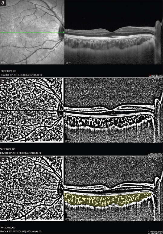
In this prospective cross-sectional study, 39 patients with unilateral anisometropic amblyopic patients and 33 eyes of 33 healthy control participants were involved. These participants were examined in terms of axial length (AL), spherical equivalent (SE), central macular thickness (CMT), choroidal thickness (CT), total choroidal area (TCA), luminal area (LA), stromal area (SA), LA/SA ratio, and CVI. All parameters were compared between amblyopic eyes, healthy fellow eyes, and healthy control eyes. The Shapiro-Wilk tests, Chi-square test, the paired t-test, Wilcoxon signed-rank test, Mann-Whitney U test, Kruskal-Wallis test, and Pearson/Spearman correlation tests were used.
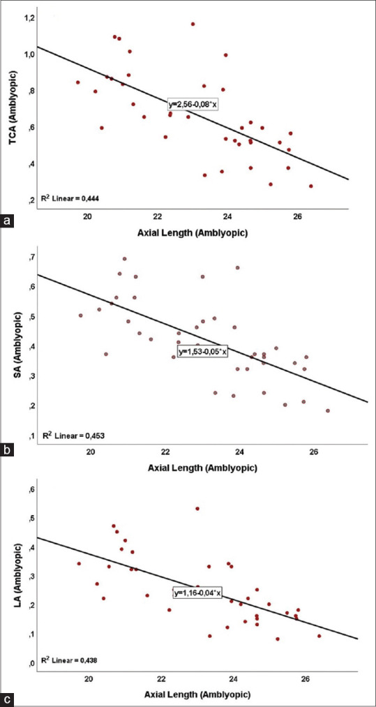
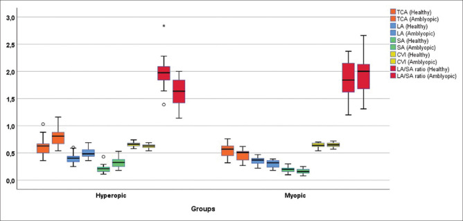
In the hyperopic patients; SE, subfoveal CT, nasal CT, temporal CT, TCA, LA, SA, and CMT were greater in amblyopic eyes than in healthy fellow eyes and control eyes (P < 0.001, P < 0.001, P < 0.001, P < 0.001, P < 0.001, P < 0.001, and P < 0.001, respectively), and CVI, LA/SA ratio, and AL were smaller in amblyopic eyes than in healthy fellow eyes and control eyes ([P < 0.001, P = 0.006], P < 0.001, and P < 0.001, respectively). In the myopic patients, subfoveal CT, nasal CT, temporal CT, TCA, LA, SA values were statistically smaller in amblyopic eyes than in healthy eyes and control eyes ([P < 0.001, P = 0.002), [P = 0.004, P = 0.012], [P = 0.012, P = 0.032], [P < 0.001, P = 0.013], [P < 0.001, P = 0.024], and [P < 0.001, P = 0.047], respectively). The differences in the AL and choroidal parameters were due to myopia and hyperopia.
The choroidal structural parameters of the amblyopic eyes were different from that of the healthy eyes.
探讨屈光参差性弱视患者脉络膜血管指数(CVI)及脉络膜形态特征。
本前瞻性病例对照研究纳入 39 例单侧屈光参差性弱视患者和 33 名健康对照者的 33 只眼。评估指标包括眼轴长度(AL)、等效球镜(SE)、中心黄斑厚度(CMT)、脉络膜厚度(CT)、脉络膜总面积(TCA)、管腔面积(LA)、基质面积(SA)、LA/SA 比值和 CVI。比较弱视眼、对侧正常眼和健康对照组的所有参数。采用 Shapiro-Wilk 检验、卡方检验、配对 t 检验、Wilcoxon 符号秩检验、Mann-Whitney U 检验、Kruskal-Wallis 检验和 Pearson/Spearman 相关检验。
远视患者中,弱视眼的 SE、黄斑中心凹下 CT、鼻侧 CT、颞侧 CT、TCA、LA、SA 和 CMT 均大于对侧正常眼和健康对照组(P<0.001,P<0.001,P<0.001,P<0.001,P<0.001,P<0.001,P<0.001),而 CVI、LA/SA 比值和 AL 则小于对侧正常眼和健康对照组(P<0.001,P=0.006)。近视患者中,弱视眼的黄斑中心凹下 CT、鼻侧 CT、颞侧 CT、TCA、LA、SA 值均小于对侧正常眼和健康对照组(P<0.001,P=0.002)。
弱视眼的脉络膜结构参数与正常眼不同。