• 文献检索
  • 文档翻译
  • 深度研究
  • 学术资讯
  • Suppr Zotero 插件Zotero 插件
  • 邀请有礼
  • 套餐&价格
  • 历史记录
应用&插件
Suppr Zotero 插件Zotero 插件浏览器插件Mac 客户端Windows 客户端微信小程序
定价
高级版会员购买积分包购买API积分包
服务
文献检索文档翻译深度研究API 文档MCP 服务
关于我们
关于 Suppr公司介绍联系我们用户协议隐私条款
关注我们

Suppr 超能文献

核心技术专利:CN118964589B侵权必究
粤ICP备2023148730 号-1Suppr @ 2026

文献检索

告别复杂PubMed语法,用中文像聊天一样搜索,搜遍4000万医学文献。AI智能推荐,让科研检索更轻松。

立即免费搜索

文件翻译

保留排版,准确专业,支持PDF/Word/PPT等文件格式,支持 12+语言互译。

免费翻译文档

深度研究

AI帮你快速写综述,25分钟生成高质量综述,智能提取关键信息,辅助科研写作。

立即免费体验

颈动脉斑块成分与无创成像在预防卒中中的重要性

Carotid Plaque Composition and the Importance of Non-Invasive in Imaging Stroke Prevention.

作者信息

Geiger Martin Andreas, Flumignan Ronald Luiz Gomes, Sobreira Marcone Lima, Avelar Wagner Mauad, Fingerhut Carla, Stein Sokrates, Guillaumon Ana Terezinha

机构信息

Division of Vascular Surgery, Department of Surgery, Universidade Estadual de Campinas-UNICAMP, São Paulo, Brazil.

Division of Vascular and Endovascular Surgery, Department of Surgery, Universidade Federal de São Paulo, São Paulo, Brazil.

出版信息

Front Cardiovasc Med. 2022 May 16;9:885483. doi: 10.3389/fcvm.2022.885483. eCollection 2022.

DOI:10.3389/fcvm.2022.885483
PMID:35651908
原文链接:https://pmc.ncbi.nlm.nih.gov/articles/PMC9149096/
Abstract

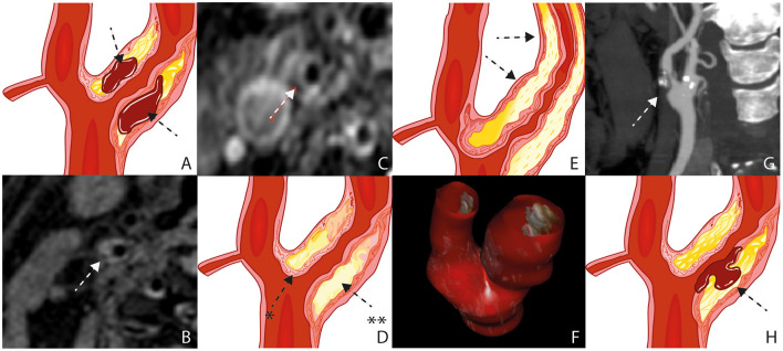
Luminal stenosis has been the standard feature for the current management strategies in patients with atherosclerotic carotid disease. Histological and imaging studies show considerable differences between plaques with identical degrees of stenosis. They indicate that specific plaque characteristics like Intraplaque hemorrhage, Lipid Rich Necrotic Core, Plaque Inflammation, Thickness and Ulceration are responsible for the increased risk of ischemic events. Intraplaque hemorrhage is defined by the accumulation of blood components within the plaque, Lipid Rich Necrotic Core is composed of macrophages loaded with lipid, Plaque Inflammation is defined as the process of atherosclerosis itself and Plaque thickness and Ulceration are defined as morphological features. Advances in imaging methods like Magnetic Resonance Imaging, Ultrasound, Computed Tomography and Positron Emission Tomography have enabled a more detailed characterization of the plaque, and its vulnerability is linked to these characteristics, changing the management of these patients based only on the degree of plaque stenosis. Studies like Rotterdam, ARIC, PARISK, CAPIAS and BIOVASC were essential to evaluate and prove the relevance of these characteristics with cerebrovascular symptoms. A better approach for the prevention of stroke is needed. This review summarizes the more frequent carotid plaque features and the available validation from recent studies with the latest evidence.

摘要

管腔狭窄一直是目前动脉粥样硬化性颈动脉疾病患者管理策略的标准特征。组织学和影像学研究表明,狭窄程度相同的斑块之间存在显著差异。这些研究表明,斑块内出血、富含脂质的坏死核心、斑块炎症、厚度和溃疡等特定斑块特征是缺血性事件风险增加的原因。斑块内出血定义为斑块内血液成分的积聚,富含脂质的坏死核心由充满脂质的巨噬细胞组成,斑块炎症定义为动脉粥样硬化本身的过程,斑块厚度和溃疡定义为形态学特征。磁共振成像、超声、计算机断层扫描和正电子发射断层扫描等成像方法的进展使得能够更详细地描述斑块特征,并且其易损性与这些特征相关联,这改变了仅基于斑块狭窄程度对这些患者的管理方式。鹿特丹研究、动脉粥样硬化风险社区研究、巴黎卒中风险研究、颈动脉斑块成像与卒中关联研究和生物血管研究等对于评估和证明这些特征与脑血管症状的相关性至关重要。需要一种更好的预防中风的方法。本综述总结了更常见的颈动脉斑块特征以及近期研究根据最新证据进行的有效验证。

https://cdn.ncbi.nlm.nih.gov/pmc/blobs/615d/9149096/1baf6acaaaf8/fcvm-09-885483-g0001.jpg

相似文献

1
Carotid Plaque Composition and the Importance of Non-Invasive in Imaging Stroke Prevention.颈动脉斑块成分与无创成像在预防卒中中的重要性
Front Cardiovasc Med. 2022 May 16;9:885483. doi: 10.3389/fcvm.2022.885483. eCollection 2022.
2
Contemporary carotid imaging: from degree of stenosis to plaque vulnerability.当代颈动脉成像:从狭窄程度到斑块易损性
J Neurosurg. 2016 Jan;124(1):27-42. doi: 10.3171/2015.1.JNS142452. Epub 2015 Jul 31.
3
Carotid Plaque Characteristics Predict Recurrent Ischemic Stroke and TIA: The PARISK (Plaque At RISK) Study.颈动脉斑块特征预测复发性缺血性中风和短暂性脑缺血发作:PARISK(风险斑块)研究。
JACC Cardiovasc Imaging. 2022 Oct;15(10):1715-1726. doi: 10.1016/j.jcmg.2022.04.003. Epub 2022 Jun 15.
4
Review of imaging biomarkers for the vulnerable carotid plaque.易损性颈动脉斑块的影像学生物标志物综述。
JVS Vasc Sci. 2021 May 15;2:149-158. doi: 10.1016/j.jvssci.2021.03.001. eCollection 2021.
5
Coexistence of Calcification, Intraplaque Hemorrhage and Lipid Core within the Asymptomatic Atherosclerotic Carotid Plaque: The Rotterdam Study.无症状性动脉粥样硬化性颈动脉斑块内钙化、斑块内出血与脂质核心的共存:鹿特丹研究
Cerebrovasc Dis. 2015;39(5-6):319-24. doi: 10.1159/000381138. Epub 2015 May 7.
6
Extracranial Vascular Disease: Carotid Stenosis and Plaque Imaging.颅外血管疾病:颈动脉狭窄和斑块成像。
Neuroimaging Clin N Am. 2021 May;31(2):157-166. doi: 10.1016/j.nic.2021.02.002.
7
Advances in Multimodality Carotid Plaque Imaging: Expert Panel Narrative Review.多模态颈动脉斑块成像进展:专家小组叙述性综述。
AJR Am J Roentgenol. 2021 Jul;217(1):16-26. doi: 10.2214/AJR.20.24869. Epub 2021 Jan 13.
8
Sex Differences in Carotid Atherosclerosis: A Systematic Review and Meta-Analysis.颈动脉粥样硬化的性别差异:系统评价和荟萃分析。
Stroke. 2023 Feb;54(2):315-326. doi: 10.1161/STROKEAHA.122.041046. Epub 2022 Nov 29.
9
MR imaging of vulnerable carotid plaque.易损性颈动脉斑块的磁共振成像
Cardiovasc Diagn Ther. 2020 Aug;10(4):1019-1031. doi: 10.21037/cdt.2020.03.12.
10
Plaque characteristics of asymptomatic carotid stenosis and risk of stroke.无症状颈动脉狭窄斑块特征与卒中风险。
Cerebrovasc Dis. 2012;34(5-6):343-50. doi: 10.1159/000343227. Epub 2012 Nov 14.

引用本文的文献

1
Carotid plaque characteristics by computed Tomography: A diagnostic accuracy systematic review.计算机断层扫描显示的颈动脉斑块特征:一项诊断准确性的系统评价
Int J Cardiol Heart Vasc. 2025 Mar 21;58:101656. doi: 10.1016/j.ijcha.2025.101656. eCollection 2025 Jun.
2
Elucidating the Relationship between Neutrophil-Lymphocyte Ratio and Plaque Composition in Patients with Drug-Eluting Stent Restenosis by Virtual Histology-Intravascular Ultrasound.通过虚拟组织学血管内超声阐明药物洗脱支架再狭窄患者中性粒细胞与淋巴细胞比值与斑块成分之间的关系。
J Cardiovasc Dev Dis. 2024 Jul 4;11(7):211. doi: 10.3390/jcdd11070211.
3
Accuracy of duplex ultrasonography versus angiotomography for the diagnosis of extracranial internal carotid stenosis.

本文引用的文献

1
Lipoprotein(a) levels and atherosclerotic plaque characteristics in the carotid artery: The Plaque at RISK (PARISK) study.脂蛋白(a)水平与颈动脉粥样硬化斑块特征:风险斑块研究(PARISK)。
Atherosclerosis. 2021 Jul;329:22-29. doi: 10.1016/j.atherosclerosis.2021.06.004. Epub 2021 Jun 11.
2
Atherosclerotic Carotid Plaque Composition and Incident Stroke and Coronary Events.动脉粥样硬化性颈动脉斑块成分与卒中及冠状动脉事件的发生
J Am Coll Cardiol. 2021 Mar 23;77(11):1426-1435. doi: 10.1016/j.jacc.2021.01.038.
3
Evolving determinants of carotid atherosclerosis vulnerability in asymptomatic patients from the MAGNETIC observational study.
双功能超声与血管造影术诊断颅外颈内动脉狭窄的准确性比较。
Rev Col Bras Cir. 2024 Jun 14;51:e20243632. doi: 10.1590/0100-6991e-20243632-en. eCollection 2024.
4
Associations between carotid plaque shape, biomechanical parameters, and ischemic stroke in mild carotid stenosis with a single plaque.轻度颈动脉狭窄伴单发斑块时颈动脉斑块形状、生物力学参数与缺血性卒中之间的关联
Ultrasonography. 2024 May;43(3):209-219. doi: 10.14366/usg.24019. Epub 2024 Apr 2.
5
Findings of Angiography and Carotid Vessel Wall Imaging Associated with Post-Procedural Clinical Events after Carotid Artery Stenting.颈动脉支架置入术后血管造影及颈动脉血管壁成像结果与术后临床事件的相关性
Neurointervention. 2024 Mar;19(1):14-23. doi: 10.5469/neuroint.2023.00486. Epub 2024 Jan 17.
6
The prevalence of extracranial carotid atherosclerosis detected via ultrasound imaging: A single-centre study in Jeddah, Saudi Arabia.经超声成像检测到的颅外颈动脉粥样硬化的患病率:沙特阿拉伯吉达的一项单中心研究。
Saudi Med J. 2024 Jan;45(1):69-73. doi: 10.15537/smj.2024.45.1.20230503.
7
Intraplaque Neovascularization, CD68+ and iNOS2+ Macrophage Infiltrate Intensity Are Associated with Atherothrombosis and Intraplaque Hemorrhage in Severe Carotid Atherosclerosis.斑块内新生血管形成、CD68+和诱导型一氧化氮合酶2+巨噬细胞浸润强度与严重颈动脉粥样硬化中的动脉粥样硬化血栓形成及斑块内出血相关。
Biomedicines. 2023 Dec 11;11(12):3275. doi: 10.3390/biomedicines11123275.
8
Contrast-Enhanced Ultrasound Feasibility in Assessing Carotid Plaque Vulnerability-Narrative Review.超声造影评估颈动脉斑块易损性的可行性——叙述性综述
J Clin Med. 2023 Oct 9;12(19):6416. doi: 10.3390/jcm12196416.
9
Novel Imaging-Based Biomarkers for Identifying Carotid Plaque Vulnerability.基于新型影像学的颈动脉斑块易损性识别生物标志物。
Biomolecules. 2023 Aug 10;13(8):1236. doi: 10.3390/biom13081236.
10
Carotid massive intraplaque hemorrhage, lipid-rich necrotic core, and heavy circumferential calcification were associated with new ipsilateral ischemic cerebral lesions after carotid artery stenting: high-resolution magnetic resonance vessel wall imaging study.颈动脉斑块内大量出血、富含脂质的坏死核心以及严重的圆周钙化与颈动脉支架置入术后同侧新发缺血性脑损伤相关:高分辨率磁共振血管壁成像研究
Cardiovasc Diagn Ther. 2023 Apr 28;13(2):355-366. doi: 10.21037/cdt-22-543. Epub 2023 Mar 1.
无症状患者颈动脉粥样硬化易损性的演变决定因素:来自 MAGNETIC 观察性研究。
Sci Rep. 2021 Jan 27;11(1):2327. doi: 10.1038/s41598-021-81247-y.
4
Advances in Multimodality Carotid Plaque Imaging: Expert Panel Narrative Review.多模态颈动脉斑块成像进展:专家小组叙述性综述。
AJR Am J Roentgenol. 2021 Jul;217(1):16-26. doi: 10.2214/AJR.20.24869. Epub 2021 Jan 13.
5
Ultrasound methods of imaging atherosclerotic plaque in carotid arteries: examinations using contrast agents.颈动脉粥样硬化斑块的超声成像方法:使用造影剂的检查
J Ultrason. 2020 Nov;20(82):e191-e200. doi: 10.15557/JoU.2020.0032. Epub 2020 Sep 28.
6
Association between Intraplaque Hemorrhage and Vascular Remodeling in Carotid Arteries: The Plaque at RISK (PARISK) Study.颈动脉斑块内出血与血管重构的相关性研究:风险斑块(PARISK)研究。
Cerebrovasc Dis. 2021;50(1):94-99. doi: 10.1159/000511935. Epub 2020 Dec 3.
7
Complicated Carotid Artery Plaques as a Cause of Cryptogenic Stroke.复杂颈动脉斑块是隐源性卒中的病因。
J Am Coll Cardiol. 2020 Nov 10;76(19):2212-2222. doi: 10.1016/j.jacc.2020.09.532.
8
Superficial Calcification With Rotund Shape Is Associated With Carotid Plaque Rupture: An Optical Coherence Tomography Study.圆形浅表钙化与颈动脉斑块破裂相关:一项光学相干断层扫描研究
Front Neurol. 2020 Sep 18;11:563334. doi: 10.3389/fneur.2020.563334. eCollection 2020.
9
MR imaging of vulnerable carotid plaque.易损性颈动脉斑块的磁共振成像
Cardiovasc Diagn Ther. 2020 Aug;10(4):1019-1031. doi: 10.21037/cdt.2020.03.12.
10
Ultrasonography of vulnerable atherosclerotic plaque in the carotid arteries: B-mode imaging.颈动脉易损动脉粥样硬化斑块的超声检查:B 型成像
J Ultrason. 2020;20(81):e135-e145. doi: 10.15557/JoU.2020.0022. Epub 2020 Jun 15.