Fedak Andrzej, Ciuk Katarzyna, Urbanik Andrzej
Department of Radiology, Jagiellonian University Medical College , Krakow , Poland.
Students' Scientific Group at the Department of Radiology, Jagiellonian University Medical College , Krakow , Poland.
J Ultrason. 2020;20(81):e135-e145. doi: 10.15557/JoU.2020.0022. Epub 2020 Jun 15.
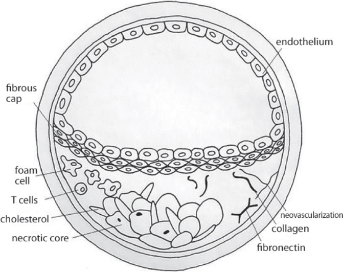
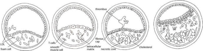
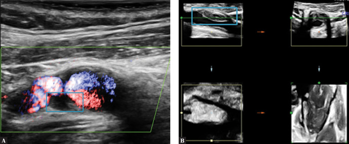
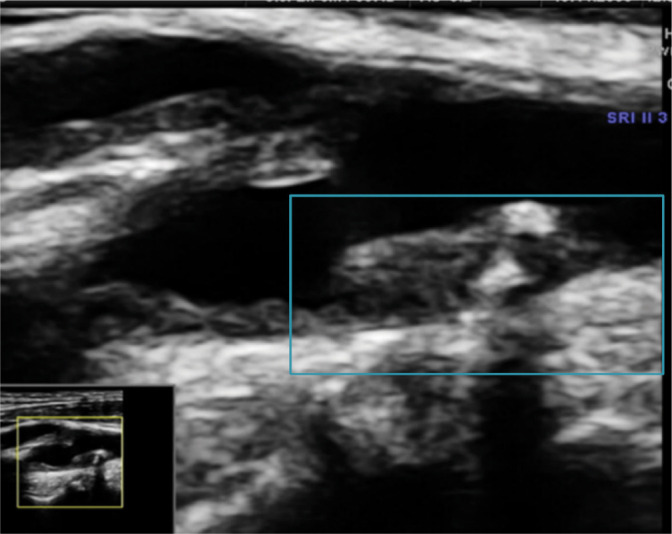
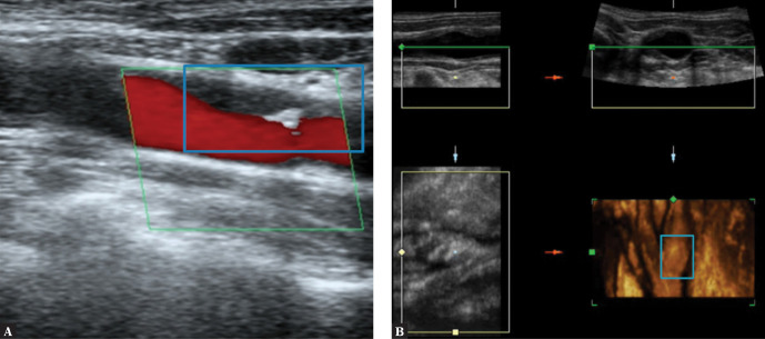
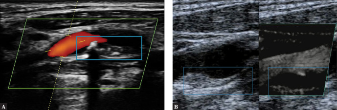
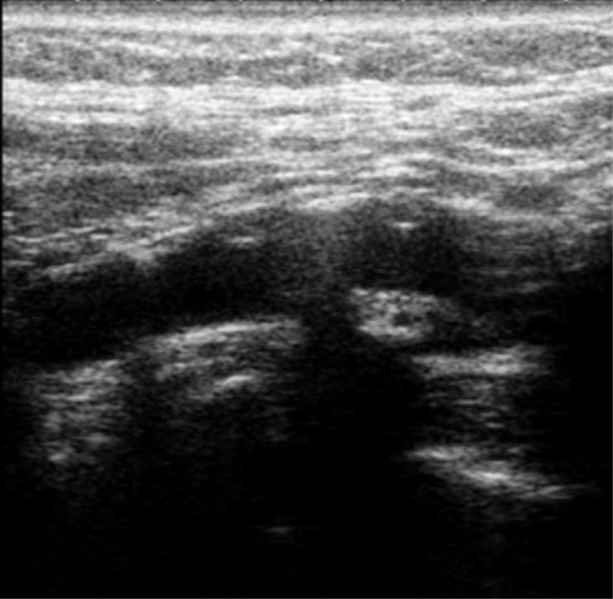
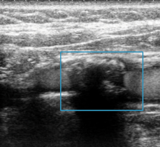
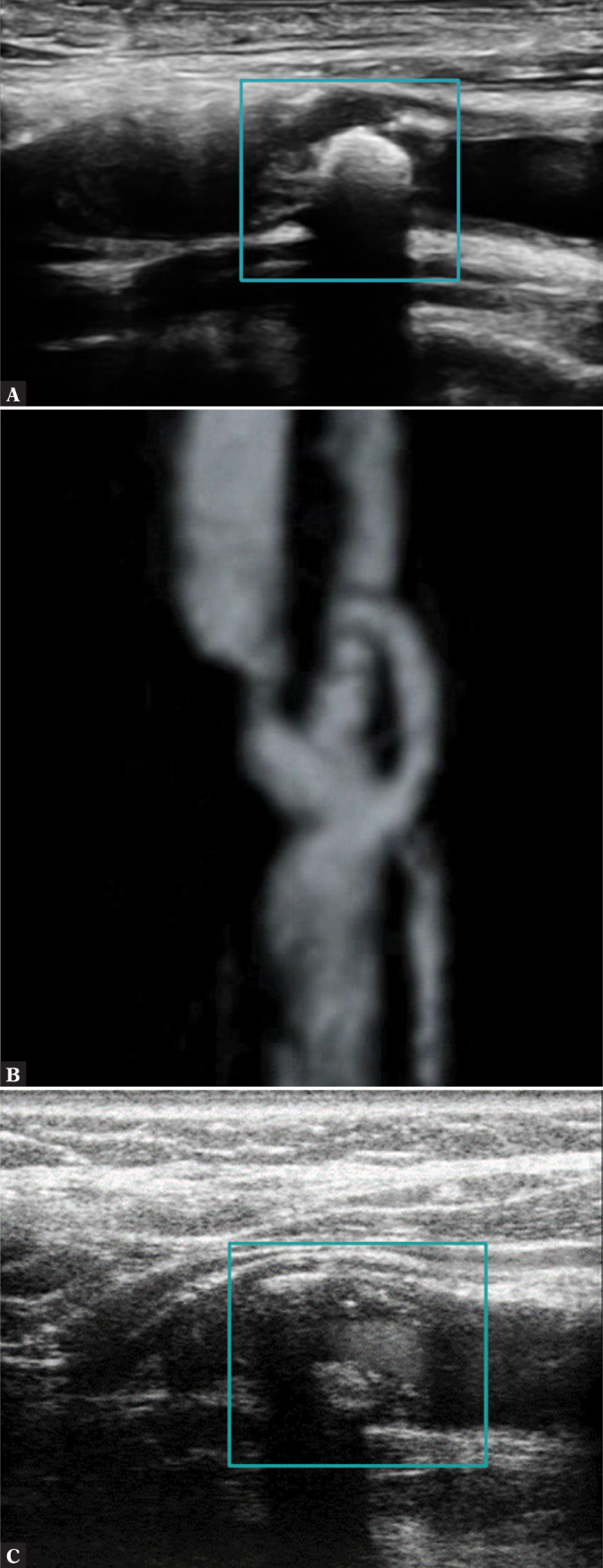
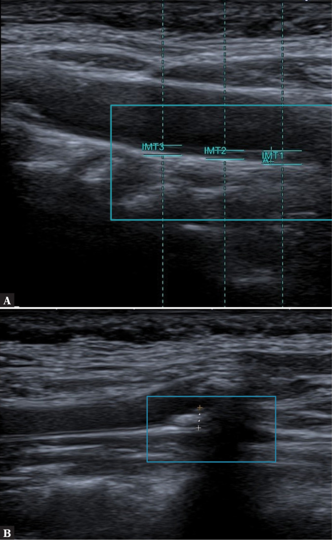
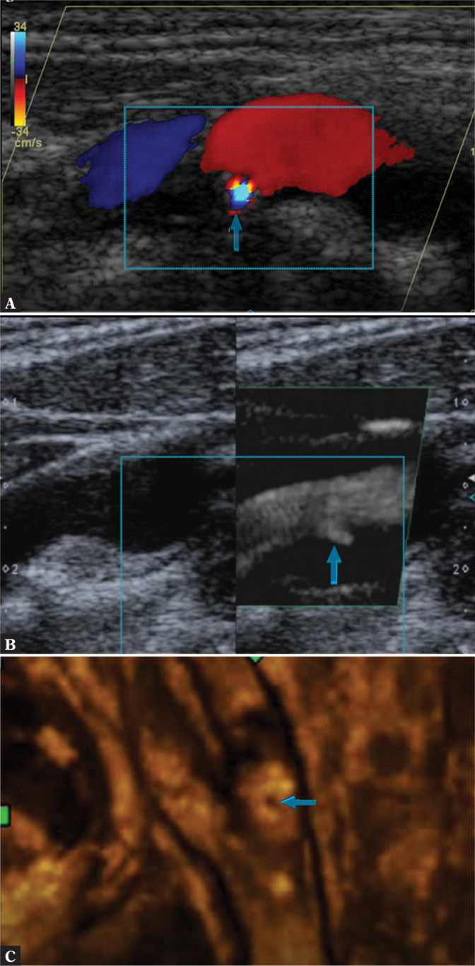
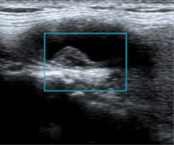
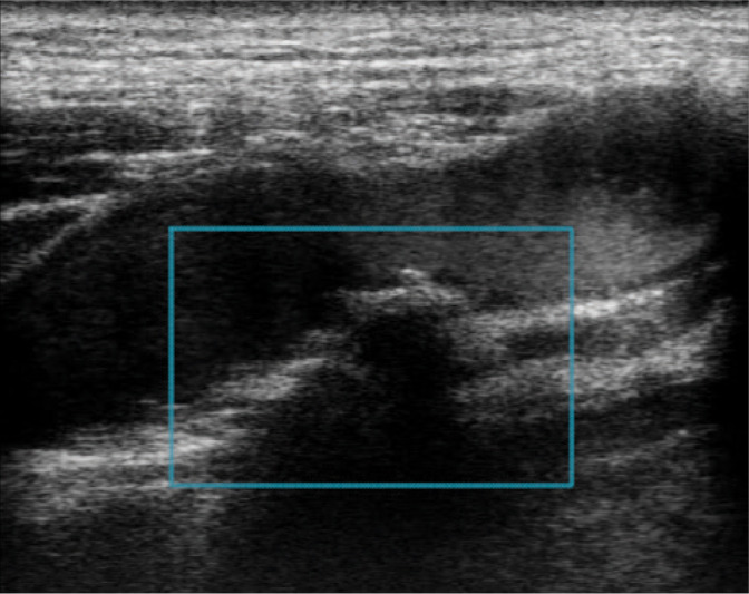
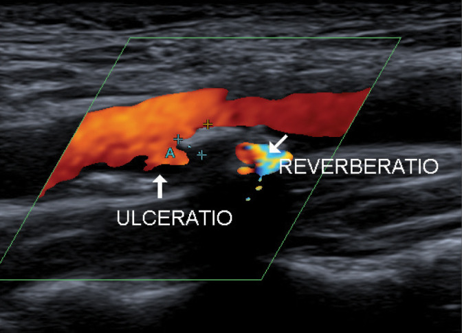
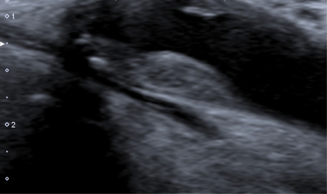
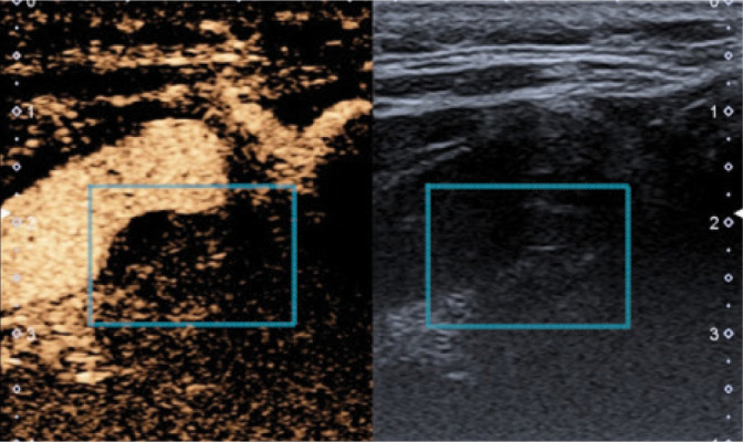
The most common type of stroke, i.e. ischemic stroke, is a great challenge for contemporary medicine as it poses both diagnostic and therapeutic difficulties. Atherosclerosis, which is rapidly beginning to affect more and more social groups, is the main cause of cerebrovascular accidents. Atherosclerosis is currently defined as a generalized, dynamic and heterogeneous inflammatory and immune process affecting arterial walls. Atherosclerotic plaque is the emanation of this disease. As the paradigm of the diagnosis of atherosclerosis has changed, it has become crucial to properly identify plaque instability within the carotid arteries by evaluating parameters and phenomena that signify a developing cascade of complications, eventually leading to stroke. Irrespective of the ultrasound technique employed, proper morphological evaluation of atherosclerotic plaque, involving observation of its echogenicity, i.e. subjective analysis of its structure, with the classification to Gray-Weale-Nicolaides types as well as assessment of the integrity of its surface, makes it possible to roughly evaluate plaque morphology and thereby its stability. This enables treatment planning and therapy monitoring. This evaluation should be a prelude to further diagnostic work-up, which involves non-invasive examinations that enable unambiguous assessment of plaque stability. These examinations include contrast-enhanced ultrasound to assess progression or recession of inflammation, which presents as plaque neovascularization, or shear wave elastography to objectively define tissue stiffness, and thereby its mineralization. The most common type of stroke, i.e. ischemic stroke, is a great challenge for contemporary medicine as it poses both diagnostic and therapeutic difficulties. Atherosclerosis, which is rapidly beginning to affect more and more social groups, is the main cause of cerebrovascular accidents. Atherosclerosis is currently defined as a generalized, dynamic and heterogeneous inflammatory and immune process affecting arterial walls. Atherosclerotic plaque is the emanation of this disease. As the paradigm of the diagnosis of atherosclerosis has changed, it has become crucial to properly identify plaque instability within the carotid arteries by evaluating parameters and phenomena that signify a developing cascade of complications, eventually leading to stroke. Irrespective of the ultrasound technique employed, proper morphological evaluation of atherosclerotic plaque, involving observation of its echogenicity, i.e. subjective analysis of its structure, with the classification to Gray-Weale–Nicolaides types as well as assessment of the integrity of its surface, makes it possible to roughly evaluate plaque morphology and thereby its stability. This enables treatment planning and therapy monitoring. This evaluation should be a prelude to further diagnostic work-up, which involves non-invasive examinations that enable unambiguous assessment of plaque stability. These examinations include contrast-enhanced ultrasound to assess progression or recession of inflammation, which presents as plaque neovascularization, or shear wave elastography to objectively define tissue stiffness, and thereby its mineralization.
最常见的中风类型,即缺血性中风,对当代医学来说是一个巨大挑战,因为它带来了诊断和治疗方面的困难。动脉粥样硬化正迅速开始影响越来越多的社会群体,它是脑血管意外的主要原因。动脉粥样硬化目前被定义为一种影响动脉壁的全身性、动态性和异质性炎症及免疫过程。动脉粥样硬化斑块是这种疾病的产物。随着动脉粥样硬化诊断模式的改变,通过评估预示着一系列并发症不断发展最终导致中风的参数和现象,来正确识别颈动脉内斑块的不稳定性变得至关重要。无论采用何种超声技术,对动脉粥样硬化斑块进行适当的形态学评估,包括观察其回声性,即对其结构进行主观分析,并分类为格雷 - 韦尔 - 尼古拉ides类型,以及评估其表面的完整性,都能够大致评估斑块形态及其稳定性。这有助于制定治疗计划和监测治疗效果。这种评估应该是进一步诊断检查的前奏,进一步诊断检查包括能够明确评估斑块稳定性的非侵入性检查。这些检查包括用于评估炎症进展或消退情况的超声造影,炎症表现为斑块新生血管形成,以及用于客观确定组织硬度从而确定其矿化程度的剪切波弹性成像。最常见的中风类型,即缺血性中风,对当代医学来说是一个巨大挑战,因为它带来了诊断和治疗方面的困难。动脉粥样硬化正迅速开始影响越来越多的社会群体,它是脑血管意外的主要原因。动脉粥样硬化目前被定义为一种影响动脉壁的全身性、动态性和异质性炎症及免疫过程。动脉粥样硬化斑块是这种疾病的产物。随着动脉粥样硬化诊断模式的改变,通过评估预示着一系列并发症不断发展最终导致中风的参数和现象,来正确识别颈动脉内斑块的不稳定性变得至关重要。无论采用何种超声技术,对动脉粥样硬化斑块进行适当的形态学评估,包括观察其回声性,即对其结构进行主观分析,并分类为格雷 - 韦尔 - 尼古拉ides类型,以及评估其表面的完整性,都能够大致评估斑块形态及其稳定性。这有助于制定治疗计划和监测治疗效果。这种评估应该是进一步诊断检查的前奏,进一步诊断检查包括能够明确评估斑块稳定性的非侵入性检查。这些检查包括用于评估炎症进展或消退情况的超声造影,炎症表现为斑块新生血管形成,以及用于客观确定组织硬度从而确定其矿化程度的剪切波弹性成像。