• 文献检索
  • 文档翻译
  • 深度研究
  • 学术资讯
  • Suppr Zotero 插件Zotero 插件
  • 邀请有礼
  • 套餐&价格
  • 历史记录
应用&插件
Suppr Zotero 插件Zotero 插件浏览器插件Mac 客户端Windows 客户端微信小程序
定价
高级版会员购买积分包购买API积分包
服务
文献检索文档翻译深度研究API 文档MCP 服务
关于我们
关于 Suppr公司介绍联系我们用户协议隐私条款
关注我们

Suppr 超能文献

核心技术专利:CN118964589B侵权必究
粤ICP备2023148730 号-1Suppr @ 2026

文献检索

告别复杂PubMed语法,用中文像聊天一样搜索,搜遍4000万医学文献。AI智能推荐,让科研检索更轻松。

立即免费搜索

文件翻译

保留排版,准确专业,支持PDF/Word/PPT等文件格式,支持 12+语言互译。

免费翻译文档

深度研究

AI帮你快速写综述,25分钟生成高质量综述,智能提取关键信息,辅助科研写作。

立即免费体验

卵巢浆液性表面乳头状交界性肿瘤的多模态超声特征

The Multimodal Ultrasound Features of Ovarian Serous Surface Papillary Borderline Tumor.

作者信息

Xie Lijun, Liu Xinxiu, Li Haiying, Huang Liyan, Chen Fang, Wang Xingfu, Jiang Lei, Gan Ling

机构信息

Department of Medical Ultrasound, The First Affiliated Hospital of Fujian Medical University, Fuzhou, China.

Department of Medical Pathology, The First Affiliated Hospital of Fujian Medical University, Fuzhou, China.

出版信息

Womens Health Rep (New Rochelle). 2022 May 10;3(1):523-532. doi: 10.1089/whr.2021.0140. eCollection 2022.

DOI:10.1089/whr.2021.0140
PMID:35652002
原文链接:https://pmc.ncbi.nlm.nih.gov/articles/PMC9148660/
Abstract

AIM

Ovarian serous surface papillary borderline tumor (OSSPBT) is very rare. Combined with clinical and pathological features, we aim to investigate the multimodal ultrasound features of OSSPBT.

PATIENTS AND METHODS

There were only 18 patients diagnosed with OSSPBT among the 142 patients who were diagnosed with borderline serous ovarian tumor by pathology from June 2008 to December 2020 in our hospital. Their clinical data, conventional ultrasound, two-dimensional contrast-enhanced ultrasound (2D-CEUS), three-dimensional contrast-enhanced ultrasound (3D-CEUS) characteristics, pathology, and prognosis were retrospectively analyzed.

RESULTS

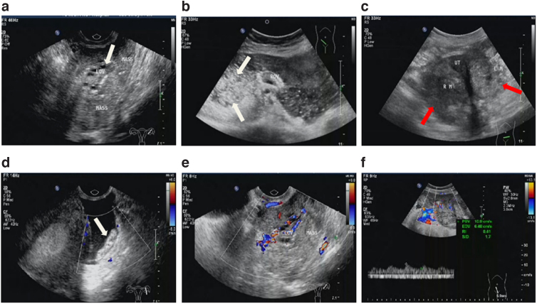
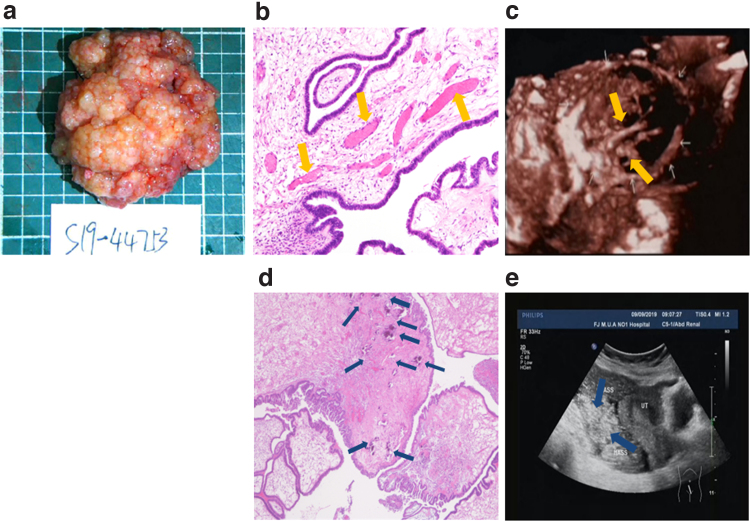
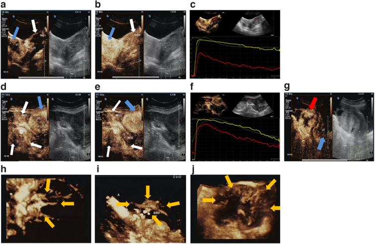
The 18 patients had no specific clinical symptoms. Multiple implantable nodules were found in 8 patients (44.4%), ascites in 13 patients (72.2%), and elevated carbohydrate antigen 125 (CA125) in 15 patients (83.3%). After excluding 2 misdiagnosed patients from 18 patients, 26 tumors in 16 patients (6 unilateral and 10 bilateral) were studied. Conventional ultrasound findings of OSSPBT showed that large solid masses around normal ovary without capsule, and numerous small dense anechoic areas were observed in the parenchyma of the lesion, with strong speckle echo ("blizzard" sign) of varying degrees. The 2D-CEUS and 3D-CEUS showed a normal ovary in the center surrounded by a radial blood supply of OSSPBT with thick and irregular branches. Histopathologically, the papillary fibrous stalk of OSSPBT had a large number of sand bodies and tortuous dilated microvessels. All patients had no recurrence after surgery, and two of them delivered successfully through assisted reproductive technology.

CONCLUSION

OSSPBT has a good prognosis. Its conventional ultrasound is characterized by irregular solid masses surrounding normal ovaries and a large number of "blizzard" signs. It showed low enhancement of eccentricity with irregular radial branches centered on the ovary by CEUS.

摘要

目的

卵巢浆液性表面乳头状交界性肿瘤(OSSPBT)非常罕见。结合临床和病理特征,我们旨在研究OSSPBT的多模态超声特征。

患者与方法

2008年6月至2020年12月在我院经病理诊断为浆液性卵巢交界性肿瘤的142例患者中,仅18例被诊断为OSSPBT。对其临床资料、常规超声、二维对比增强超声(2D-CEUS)、三维对比增强超声(3D-CEUS)特征、病理及预后进行回顾性分析。

结果

18例患者无特异性临床症状。8例患者(44.4%)发现多发可植入结节,13例患者(72.2%)有腹水,15例患者(83.3%)糖类抗原125(CA125)升高。18例患者中排除2例误诊患者后,对16例患者(6例单侧和10例双侧)的26个肿瘤进行研究。OSSPBT的常规超声表现为正常卵巢周围有大的实性肿块,无包膜,病变实质内可见大量小的密集无回声区,伴有不同程度的强斑点状回声(“暴风雪”征)。2D-CEUS和3D-CEUS显示中心为正常卵巢,周围为OSSPBT的放射状血供,分支粗大且不规则。组织病理学上,OSSPBT的乳头状纤维蒂有大量砂粒体和迂曲扩张的微血管。所有患者术后均无复发,其中2例通过辅助生殖技术成功分娩。

结论

OSSPBT预后良好。其常规超声特征为正常卵巢周围有不规则实性肿块及大量“暴风雪”征。CEUS显示以卵巢为中心的偏心性低增强,有不规则放射状分支。

https://cdn.ncbi.nlm.nih.gov/pmc/blobs/7b4c/9148660/0321ed0414e8/whr.2021.0140_figure3.jpg
https://cdn.ncbi.nlm.nih.gov/pmc/blobs/7b4c/9148660/13046baa3e02/whr.2021.0140_figure1.jpg
https://cdn.ncbi.nlm.nih.gov/pmc/blobs/7b4c/9148660/8fad53f26f3f/whr.2021.0140_figure2.jpg
https://cdn.ncbi.nlm.nih.gov/pmc/blobs/7b4c/9148660/0321ed0414e8/whr.2021.0140_figure3.jpg
https://cdn.ncbi.nlm.nih.gov/pmc/blobs/7b4c/9148660/13046baa3e02/whr.2021.0140_figure1.jpg
https://cdn.ncbi.nlm.nih.gov/pmc/blobs/7b4c/9148660/8fad53f26f3f/whr.2021.0140_figure2.jpg
https://cdn.ncbi.nlm.nih.gov/pmc/blobs/7b4c/9148660/0321ed0414e8/whr.2021.0140_figure3.jpg

相似文献

1
The Multimodal Ultrasound Features of Ovarian Serous Surface Papillary Borderline Tumor.卵巢浆液性表面乳头状交界性肿瘤的多模态超声特征
Womens Health Rep (New Rochelle). 2022 May 10;3(1):523-532. doi: 10.1089/whr.2021.0140. eCollection 2022.
2
Ovarian serous surface papillary borderline tumor: characteristic imaging features with clinicopathological correlation.卵巢浆液性表面乳头状交界性肿瘤:与临床病理相关性的特征性影像学表现。
Br J Radiol. 2018 Jul;91(1088):20170689. doi: 10.1259/bjr.20170689. Epub 2018 Jun 21.
3
Benign Brenner tumor of the ovary: two-dimensional and contrast-enhanced ultrasound features-a retrospective study from a single center.卵巢良性勃勒纳瘤:二维及对比增强超声特征——来自单一中心的回顾性研究
Front Oncol. 2024 Mar 8;14:1337806. doi: 10.3389/fonc.2024.1337806. eCollection 2024.
4
Encapsulated papillary carcinoma of breast: Comparative study of multimodal ultrasound manifestations and pathological features.乳腺包膜内乳头状癌:多模态超声表现与病理特征的对比研究
Clin Hemorheol Microcirc. 2024;88(4):537-549. doi: 10.3233/CH-242263.
5
Serous surface papillary borderline ovarian tumors: correlation of sonographic features with clinic pathological findings.浆液性表面乳头状交界性卵巢肿瘤:超声特征与临床病理结果的相关性
Ultrasound Obstet Gynecol. 2024 May;63(5):691-698. doi: 10.1002/uog.27454.
6
Ultrasonographic and clinicopathological features of pelvic yolk sac tumors in women: a single-center retrospective analysis.女性盆腔卵黄囊瘤的超声及临床病理特征:单中心回顾性分析
Front Oncol. 2024 Jun 20;14:1417761. doi: 10.3389/fonc.2024.1417761. eCollection 2024.
7
Ultrasound features and clinical outcome of patients with malignant ovarian masses diagnosed during pregnancy: experience of a gynecological oncology ultrasound center.孕期诊断为卵巢恶性肿块患者的超声特征及临床结局:妇科肿瘤超声中心的经验
Int J Gynecol Cancer. 2019 Sep;29(7):1182-1194. doi: 10.1136/ijgc-2019-000373. Epub 2019 Jul 19.
8
Ultrasound diagnosis of serous surface papillary borderline ovarian tumor: A case series with a review of the literature.浆液性表面乳头状交界性卵巢肿瘤的超声诊断:病例系列及文献复习
J Clin Ultrasound. 2015 Nov-Dec;43(9):573-7. doi: 10.1002/jcu.22266. Epub 2015 Feb 23.
9
Color Doppler ultrasound and contrast-enhanced ultrasound in the diagnosis of lacrimal apparatus tumors.彩色多普勒超声及超声造影在泪器肿瘤诊断中的应用
Oncol Lett. 2018 Aug;16(2):2215-2220. doi: 10.3892/ol.2018.8879. Epub 2018 Jun 1.
10
Combination of serum CA19-9 and CA125 levels and contrast-enhanced ultrasound parametric data facilitates to differentiate ovarian serous carcinoma from ovarian malignant epithelial cancer.血清CA19-9和CA125水平与超声造影参数数据相结合有助于鉴别卵巢浆液性癌与卵巢恶性上皮性癌。
Medicine (Baltimore). 2018 Apr;97(16):e0358. doi: 10.1097/MD.0000000000010358.

引用本文的文献

1
Ultrasonographic and clinicopathological features of pelvic yolk sac tumors in women: a single-center retrospective analysis.女性盆腔卵黄囊瘤的超声及临床病理特征:单中心回顾性分析
Front Oncol. 2024 Jun 20;14:1417761. doi: 10.3389/fonc.2024.1417761. eCollection 2024.

本文引用的文献

1
Borderline epithelial ovarian tumors: what the radiologist should know.卵巢交界性上皮性肿瘤:放射科医生应了解的内容。
Abdom Radiol (NY). 2021 Jun;46(6):2350-2366. doi: 10.1007/s00261-020-02688-z. Epub 2020 Aug 29.
2
Cerebrospinal Fluid MicroRNA Signatures as Diagnostic Biomarkers in Brain Tumors.脑脊液微小RNA特征作为脑肿瘤的诊断生物标志物
Cancers (Basel). 2019 Oct 12;11(10):1546. doi: 10.3390/cancers11101546.
3
ESMO-ESGO consensus conference recommendations on ovarian cancer: pathology and molecular biology, early and advanced stages, borderline tumours and recurrent disease.
欧洲肿瘤内科学会(ESMO)-欧洲妇科肿瘤学会(ESGO)关于卵巢癌的共识会议建议:病理学与分子生物学、早期和晚期阶段、交界性肿瘤及复发性疾病
Int J Gynecol Cancer. 2019 May 7;29(4):728-760. doi: 10.1136/ijgc-2019-000308.
4
[Perfusion Imaging of Ovarian Masses with Contrast-enhanced Ultrasonography].[超声造影对卵巢肿块的灌注成像]
Sichuan Da Xue Xue Bao Yi Xue Ban. 2018 Jul;49(4):587-593.
5
Bilateral serous surface papillary borderline ovarian tumor in 19-year-old patient. Ultrasound, immunohistochemical and therapeutic particularities of reproductive age.19岁患者的双侧浆液性表面乳头状交界性卵巢肿瘤。生殖年龄的超声、免疫组化及治疗特点。
Rom J Morphol Embryol. 2017;58(3):989-995.
6
Elevated Preoperative CA125 or CA19-9 in Borderline Ovarian Tumors: Could It Be Suggestive of Advanced Stage or a Poor Prognosis?交界性卵巢肿瘤患者术前CA125或CA19-9升高:这是否提示晚期或预后不良?
Gynecol Obstet Invest. 2018;83(1):45-51. doi: 10.1159/000475817. Epub 2017 Jun 2.
7
Imaging in gynecological disease (12): clinical and ultrasound features of invasive and non-invasive malignant serous ovarian tumors.妇科疾病影像学(十二):侵袭性和非侵袭性恶性浆液性卵巢肿瘤的临床和超声特征。
Ultrasound Obstet Gynecol. 2017 Dec;50(6):788-799. doi: 10.1002/uog.17414. Epub 2017 Nov 2.
8
Ultrasound diagnosis of serous surface papillary borderline ovarian tumor: A case series with a review of the literature.浆液性表面乳头状交界性卵巢肿瘤的超声诊断:病例系列及文献复习
J Clin Ultrasound. 2015 Nov-Dec;43(9):573-7. doi: 10.1002/jcu.22266. Epub 2015 Feb 23.
9
Long-term follow-up of borderline ovarian tumors clinical outcome and prognostic factors.卵巢交界性肿瘤的长期随访:临床结局及预后因素
Anticancer Res. 2014 Nov;34(11):6725-30.
10
Combination of 2- and 3-dimensional contrast-enhanced transvaginal sonography for diagnosis of small adnexal masses.二维和三维超声造影经阴道超声检查联合用于诊断附件小肿块
J Ultrasound Med. 2014 Nov;33(11):1889-99. doi: 10.7863/ultra.33.11.1889.