Department of Microbiology, Immunology, and Molecular Genetics, University of California, Los Angeles, CA, USA.
California NanoSystems Institute, University of California, Los Angeles, CA, USA.
Nat Struct Mol Biol. 2022 Jul;29(7):698-705. doi: 10.1038/s41594-022-00779-7. Epub 2022 Jun 2.
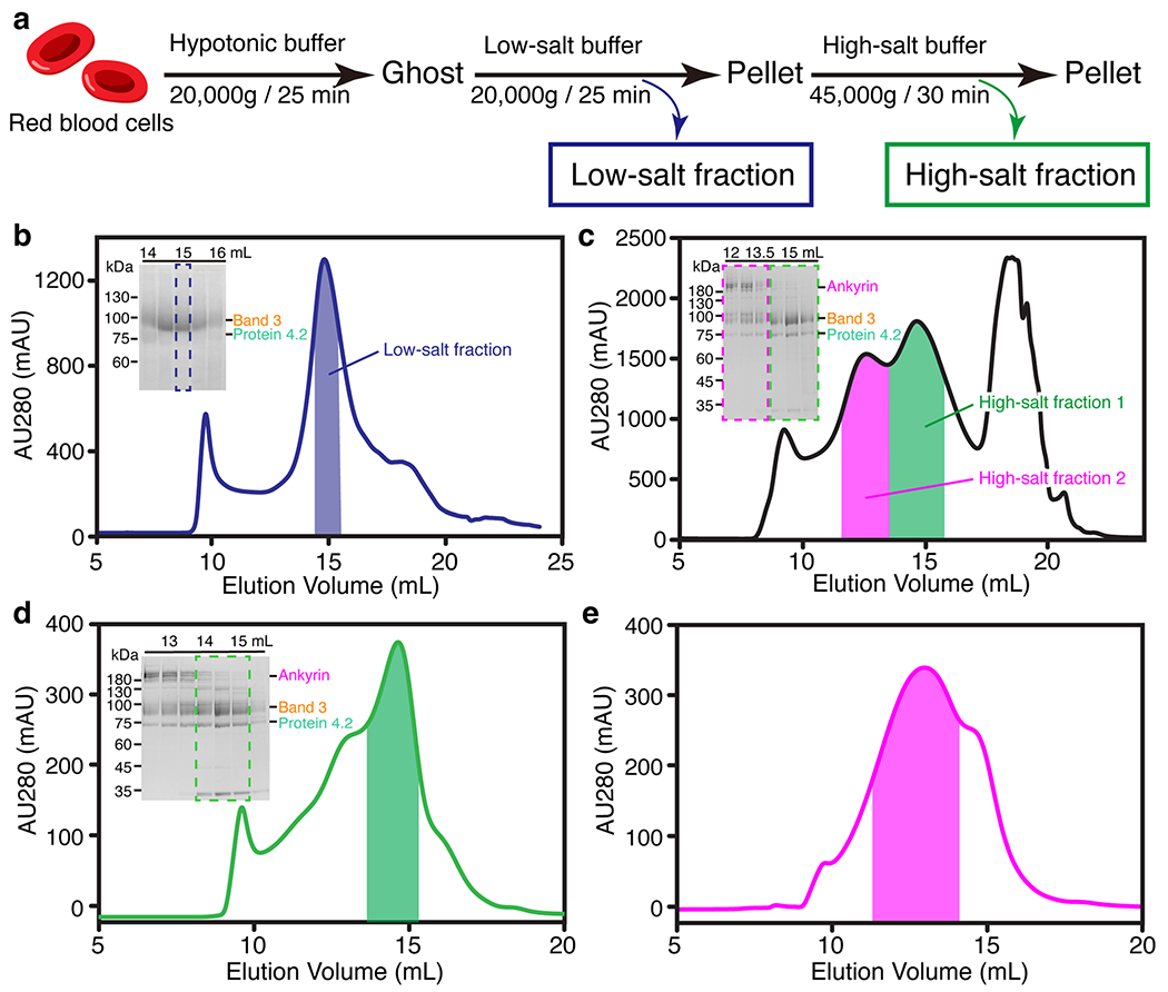
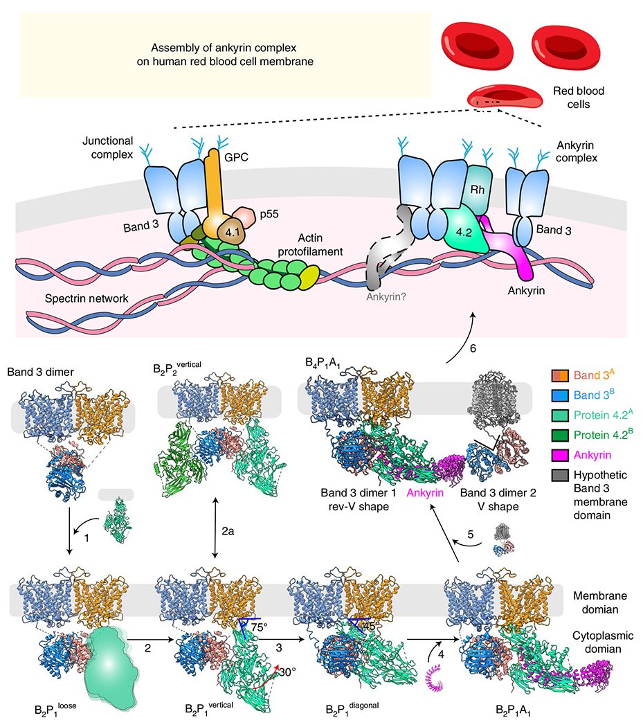
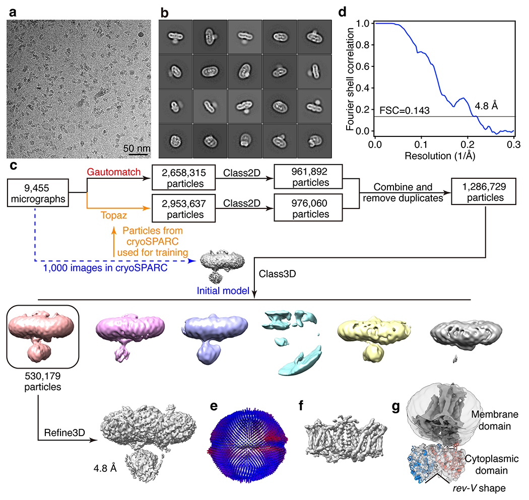
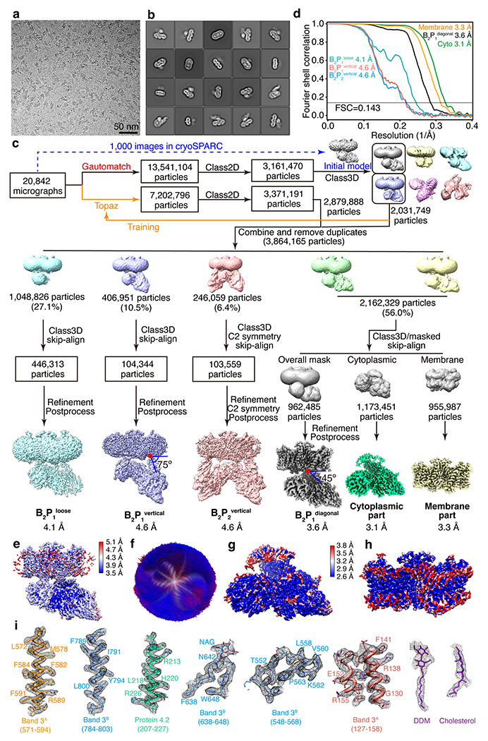
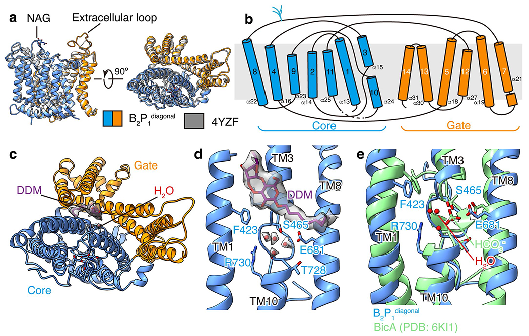
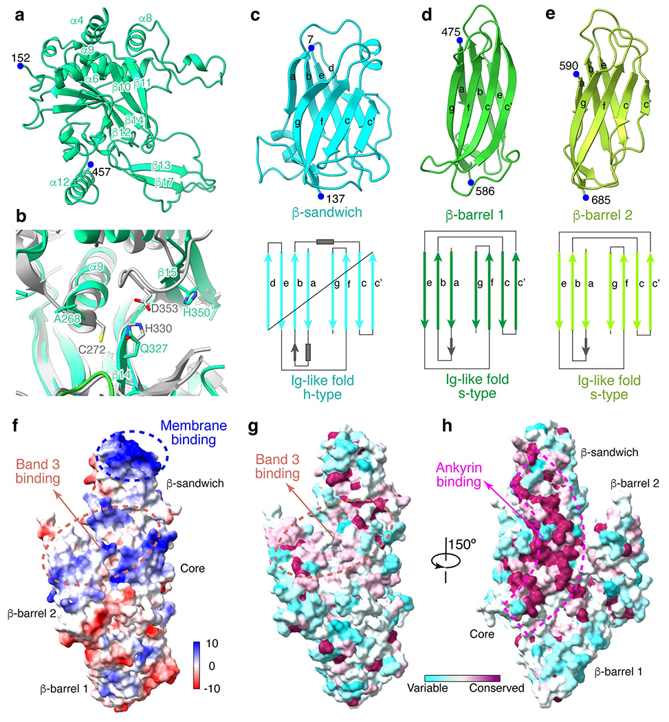
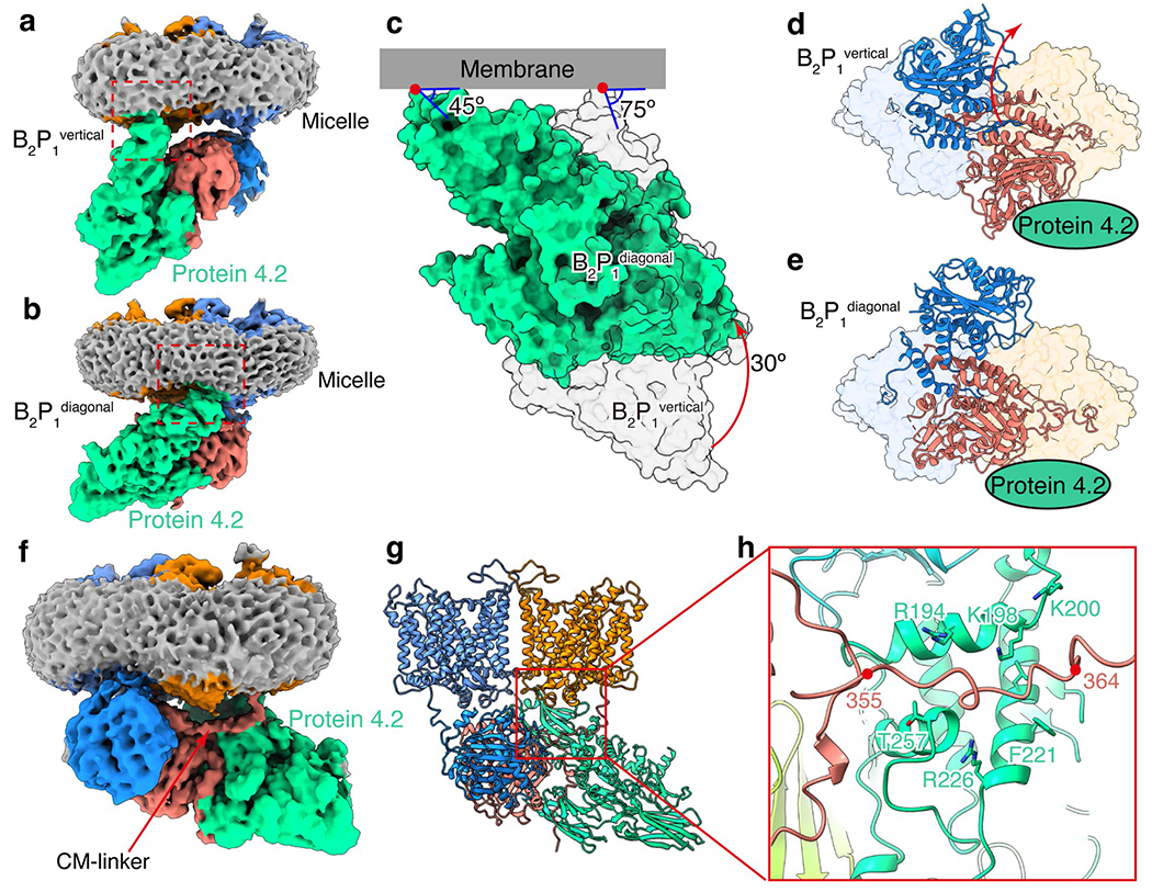
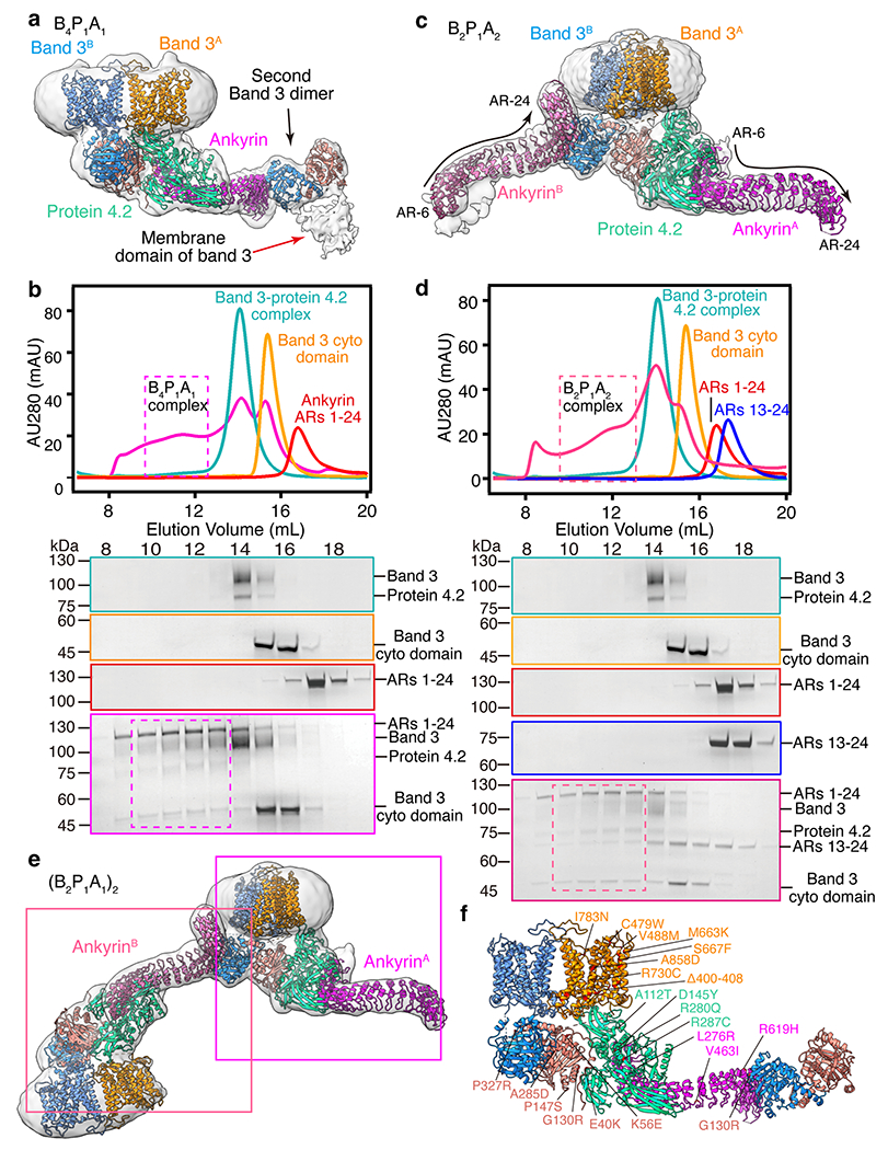
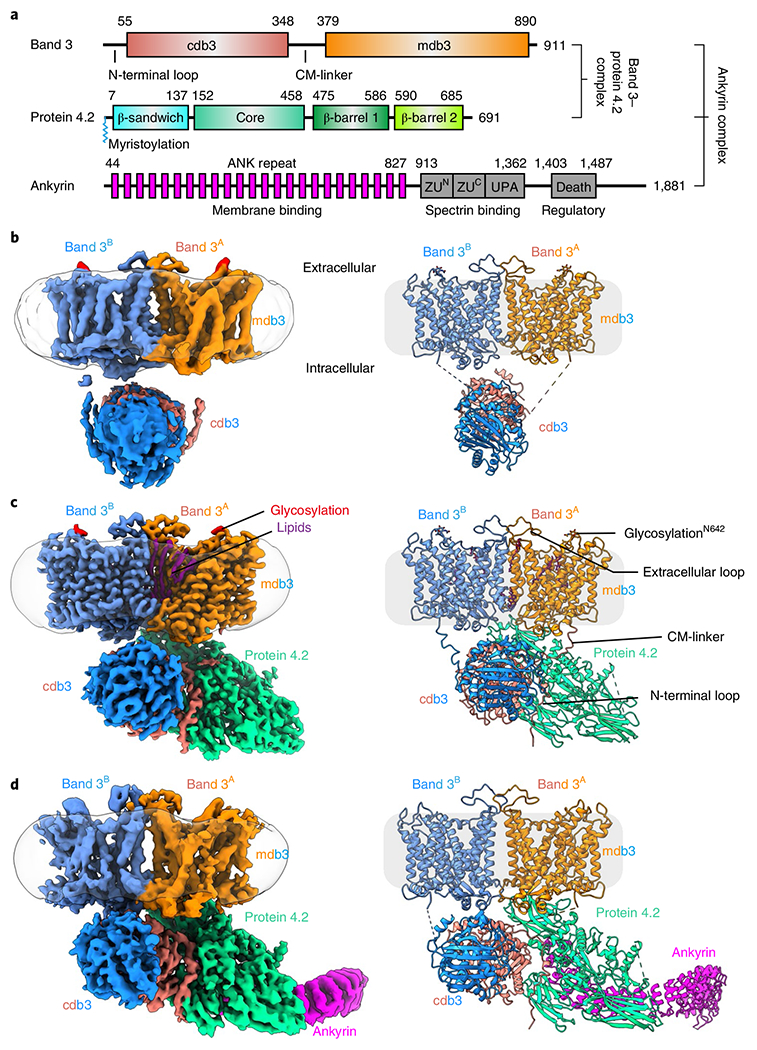
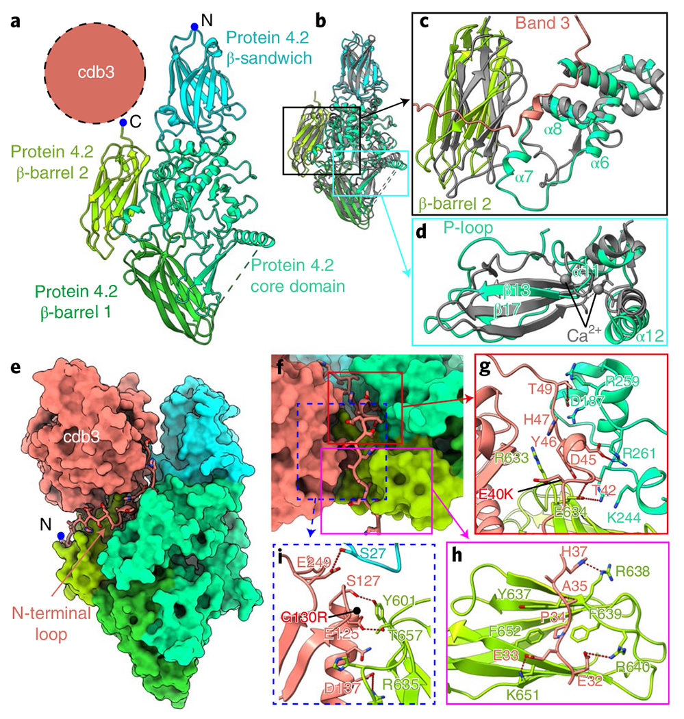
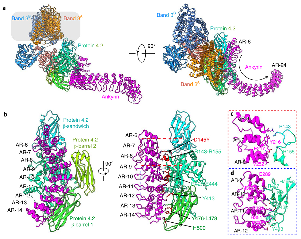
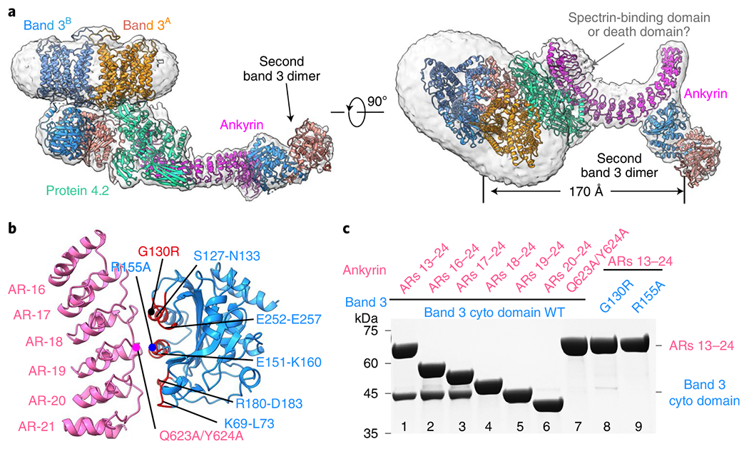
The cytoskeleton of a red blood cell (RBC) is anchored to the cell membrane by the ankyrin complex. This complex is assembled during RBC genesis and comprises primarily band 3, protein 4.2 and ankyrin, whose mutations contribute to numerous human inherited diseases. High-resolution structures of the ankyrin complex have been long sought-after to understand its assembly and disease-causing mutations. Here, we analyzed native complexes on the human RBC membrane by stepwise fractionation. Cryo-electron microscopy structures of nine band-3-associated complexes reveal that protein 4.2 stabilizes the cytoplasmic domain of band 3 dimer. In turn, the superhelix-shaped ankyrin binds to this protein 4.2 via ankyrin repeats (ARs) 6-13 and to another band 3 dimer via ARs 17-20, bridging two band 3 dimers in the ankyrin complex. Integration of these structures with both prior data and our biochemical data supports a model of ankyrin complex assembly during erythropoiesis and identifies interactions essential for the mechanical stability of RBC.
红细胞(RBC)的细胞骨架通过锚蛋白复合物固定在细胞膜上。该复合物在 RBC 生成过程中组装,主要由带 3 蛋白、蛋白 4.2 和锚蛋白组成,其突变会导致许多人类遗传性疾病。为了了解其组装和致病突变,人们长期以来一直寻求高分辨率的锚蛋白复合物结构。在这里,我们通过逐步分级分离分析了人 RBC 膜上的天然复合物。对九个与带 3 相关的复合物的冷冻电镜结构分析表明,蛋白 4.2 稳定了带 3 二聚体的细胞质结构域。反过来,超螺旋状的锚蛋白通过 ARs 6-13 与该蛋白 4.2 结合,并通过 ARs 17-20 与另一个带 3 二聚体结合,在锚蛋白复合物中桥接两个带 3 二聚体。将这些结构与之前的数据和我们的生化数据整合在一起,支持了红细胞生成过程中锚蛋白复合物组装的模型,并确定了对 RBC 机械稳定性至关重要的相互作用。