Clinical department for abdominal and general surgery, University Medical Centre Maribor, Maribor, Slovenia.
Institute of Physiology, Faculty of Medicine, University of Maribor, Maribor, Slovenia.
PLoS One. 2022 Jun 3;17(6):e0268644. doi: 10.1371/journal.pone.0268644. eCollection 2022.
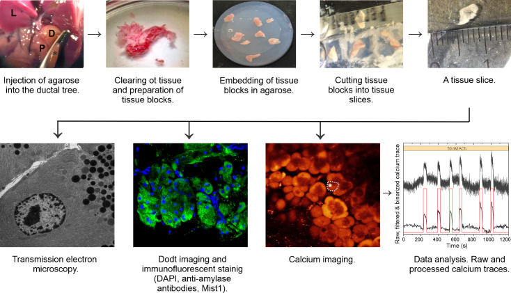
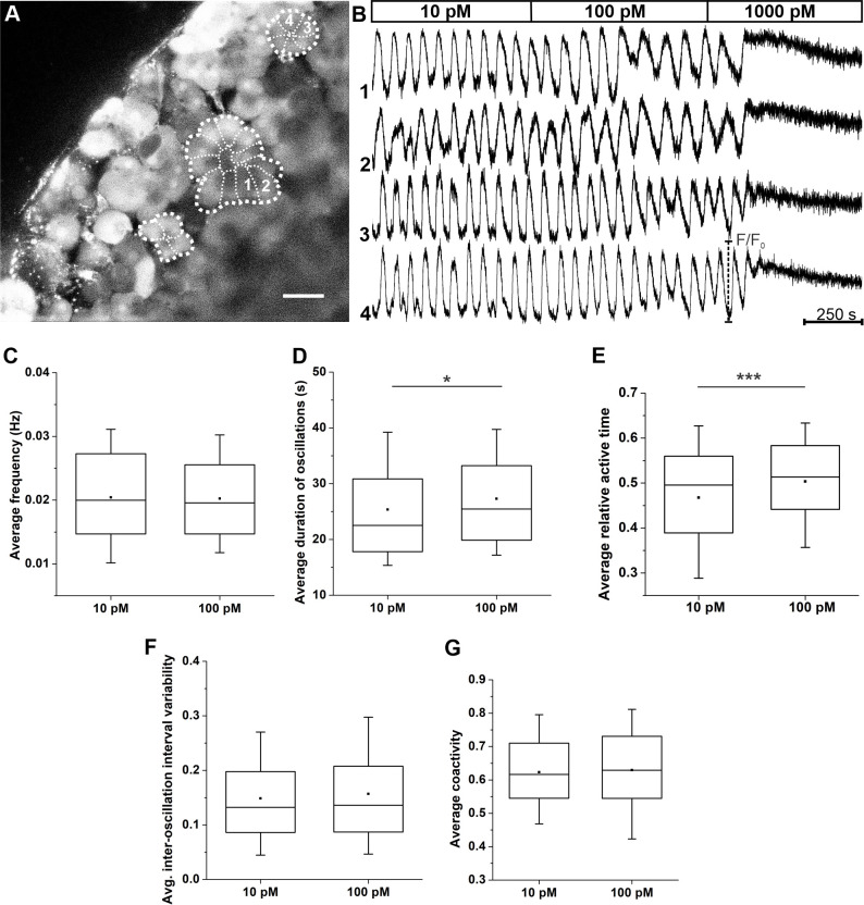
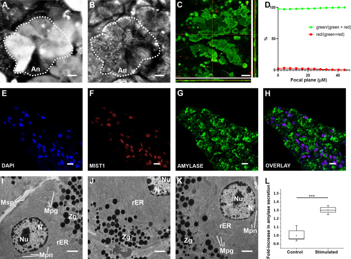
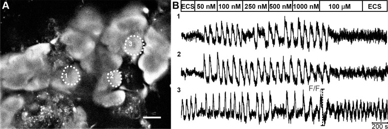
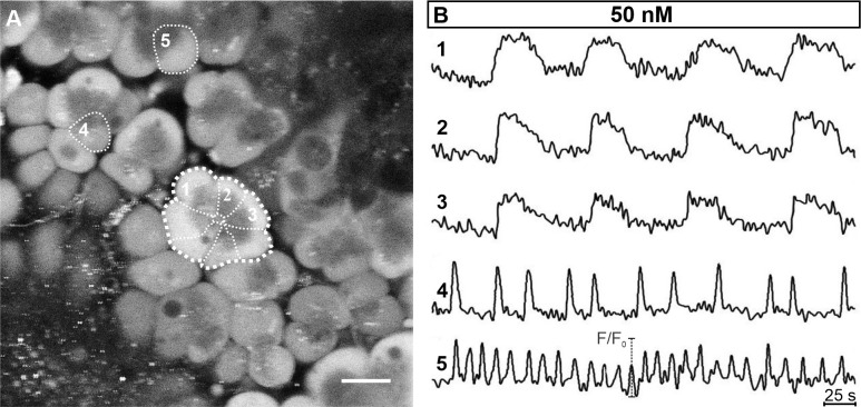
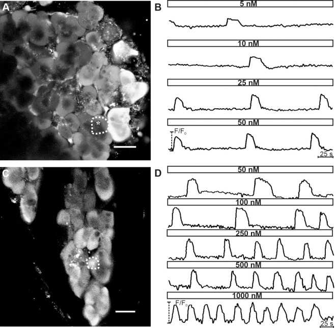
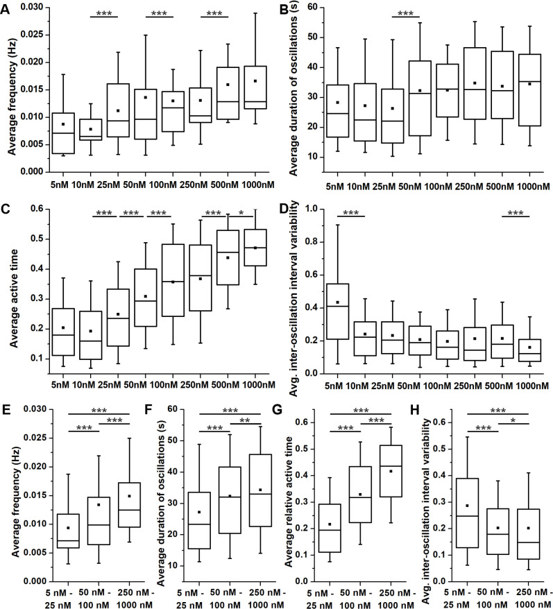
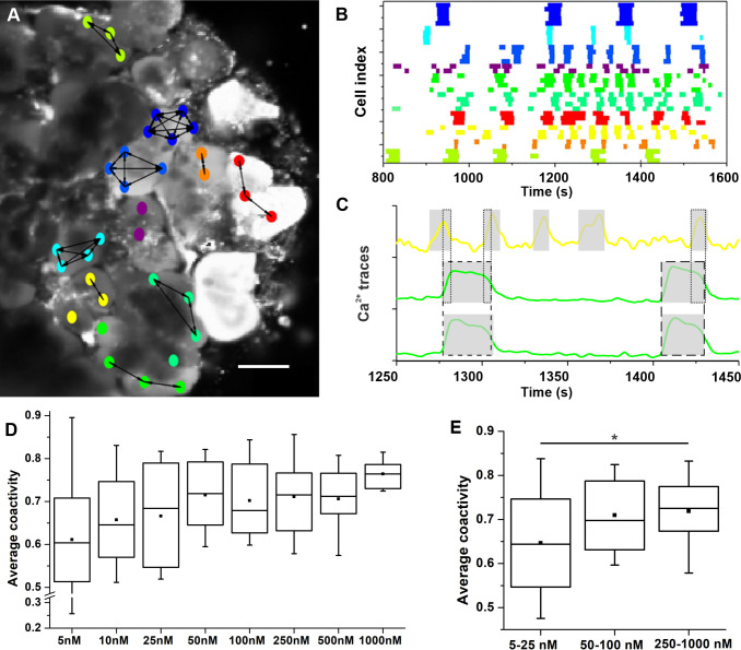
The physiology and pathophysiology of the exocrine pancreas are in close connection to changes in intra-cellular Ca2+ concentration. Most of our knowledge is based on in vitro experiments on acinar cells or acini enzymatically isolated from their surroundings, which can alter their structure, physiology, and limit our understanding. Due to these limitations, the acute pancreas tissue slice technique was introduced almost two decades ago as a complementary approach to assess the morphology and physiology of both the endocrine and exocrine pancreas in a more conserved in situ setting. In this study, we extend previous work to functional multicellular calcium imaging on acinar cells in tissue slices. The viability and morphological characteristics of acinar cells within the tissue slice were assessed using the LIVE/DEAD assay, transmission electron microscopy, and immunofluorescence imaging. The main aim of our study was to characterize the responses of acinar cells to stimulation with acetylcholine and compare them with responses to cerulein in pancreatic tissue slices, with special emphasis on inter-cellular and inter-acinar heterogeneity and coupling. To this end, calcium imaging was performed employing confocal microscopy during stimulation with a wide range of acetylcholine concentrations and selected concentrations of cerulein. We show that various calcium oscillation parameters depend monotonically on the stimulus concentration and that the activity is rather well synchronized within acini, but not between acini. The acute pancreas tissue slice represents a viable and reliable experimental approach for the evaluation of both intra- and inter-cellular signaling characteristics of acinar cell calcium dynamics. It can be utilized to assess many cells simultaneously with a high spatiotemporal resolution, thus providing an efficient and high-yield platform for future studies of normal acinar cell biology, pathophysiology, and screening pharmacological substances.
外分泌胰腺的生理学和病理生理学与细胞内钙离子浓度的变化密切相关。我们的大部分知识都是基于对从周围环境中分离出来的胰腺腺泡细胞或腺泡进行的体外实验,这些实验可能会改变它们的结构、生理学,从而限制我们的理解。由于这些限制,近二十年前引入了急性胰腺组织切片技术,作为一种补充方法,以在更保守的原位环境中评估内分泌和外分泌胰腺的形态和生理学。在这项研究中,我们将以前的工作扩展到组织切片中胰腺腺泡细胞的功能多细胞钙成像。使用 LIVE/DEAD 测定法、透射电子显微镜和免疫荧光成像评估组织切片中腺泡细胞的活力和形态特征。我们研究的主要目的是描述乙酰胆碱刺激下腺泡细胞的反应,并将其与胰腺组织切片中对 cerulein 的反应进行比较,特别强调细胞间和细胞间异质性和偶联。为此,在使用广泛的乙酰胆碱浓度和选定浓度的 cerulein 刺激期间,通过共聚焦显微镜进行钙成像。我们表明,各种钙振荡参数与刺激浓度单调相关,并且在腺泡内活动相当同步,但在腺泡之间不同步。急性胰腺组织切片代表了一种可行且可靠的实验方法,可用于评估腺泡细胞钙动力学的细胞内和细胞间信号特征。它可以用于同时评估许多细胞,具有高时空分辨率,从而为正常腺泡细胞生物学、病理生理学和筛选药物物质的未来研究提供高效、高产的平台。