Multiple Sclerosis Centre, Neurology Clinic, Department of Neuroscience, Università degli Studi di Padova, Padova, Italy.
Ophthalmology Clinic, Department of Neuroscience, Università degli Studi di Padova, Padova, Italy.
Front Immunol. 2022 May 19;13:852183. doi: 10.3389/fimmu.2022.852183. eCollection 2022.
Increasing evidence suggests that retinal hyper-reflecting foci (HRF) might be clusters of activated and proliferating microglia. Since microglia are widespread activated in multiple sclerosis (MS) brain, its evaluation in retina may help to understand and monitor MS-related pathology.
This study aims at investigating the association of HRF with cerebrospinal fluid (CSF) cytokines and MRI parameters in relapsing-remitting MS (RRMS).
Nineteen RRMS at clinical onset and 15 non-inflammatory neurological disorders (NIND) underwent brain 3T MRI and CSF examination. Optical coherence tomography (OCT) analysis, including HRF count, was performed on RRMS patients. Sixty-nine cytokines/chemokines were analyzed in the CSF by multiplex technology.
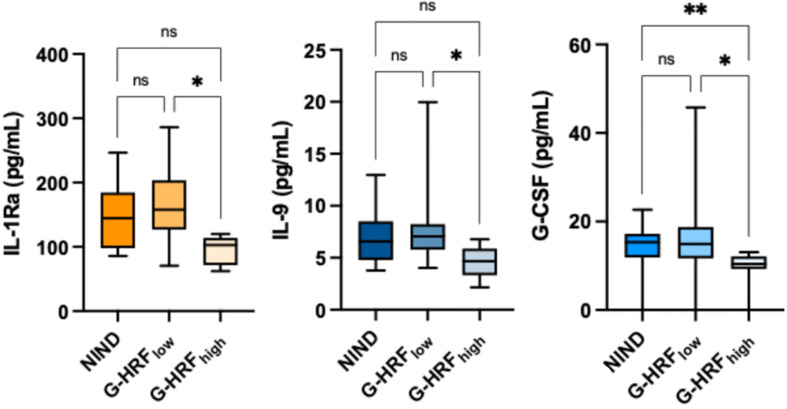
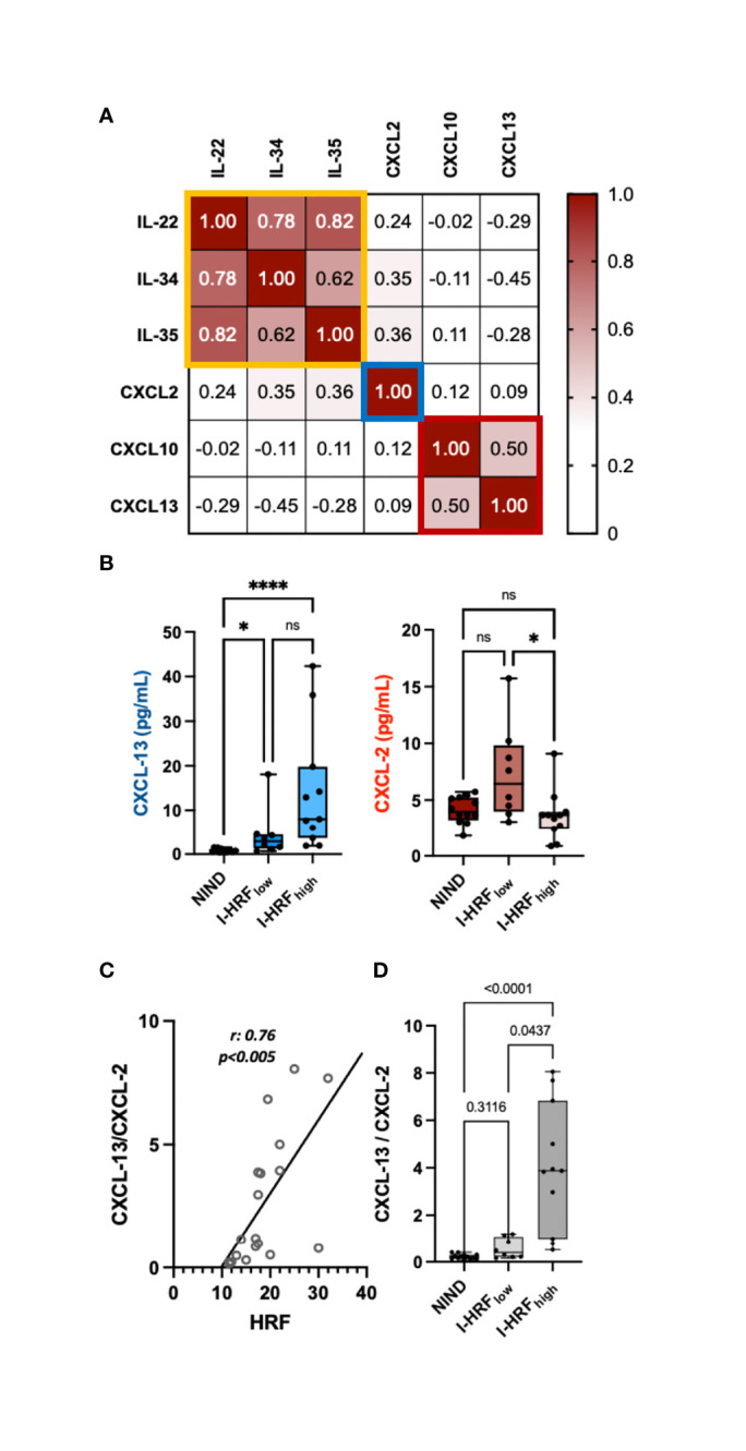
In RRMS, HRF count in the ganglion cell layer (GCL) was associated with IL-1Ra, IL-9, IL-15, IFN-γ, and G-CSF. Moreover, in RRMS patients CSF concentrations of IL-1Ra and G-CSF associated with global cortical thickness. The HRF count in the inner nuclear layer (INL) correlated with IL-22, IL-34, IL-35, CXCL-2, CXCL-10, and CXCL-13, and multivariate analysis confirmed a strong association (r: 0.47) with both CXCL-2 (β: -0.965, p = 0.0052) and CXCL-13 (β: 0.241, p = 0.018). This latter cytokine increased in RRMS with high HRF count compared with NIND and RRMS with low HRF count. Finally, the CXCL-13/CXCL-2 ratio strongly associated with HRF count (r: 0.8, p < 0.005) and cortical lesion volume (r: 0.5, p < 0.05).
The association of HRF with intrathecally produced monocyte/microglia-derived cytokines confirms their microglial origin and indicates they are worth further evaluating as markers of activated microglia.
越来越多的证据表明,视网膜高反射灶(HRF)可能是激活和增殖的小胶质细胞簇。由于小胶质细胞在多发性硬化症(MS)大脑中广泛激活,因此评估其在视网膜中的存在可能有助于了解和监测与 MS 相关的病理学。
本研究旨在探讨 HRF 与复发缓解型多发性硬化症(RRMS)患者脑脊液(CSF)细胞因子和 MRI 参数之间的关系。
19 例 RRMS 患者在发病时和 15 例非炎症性神经疾病(NIND)患者接受了脑部 3T MRI 和 CSF 检查。对 RRMS 患者进行光学相干断层扫描(OCT)分析,包括 HRF 计数。采用多重技术分析 CSF 中的 69 种细胞因子/趋化因子。
在 RRMS 患者中,神经节细胞层(GCL)的 HRF 计数与 IL-1Ra、IL-9、IL-15、IFN-γ 和 G-CSF 相关。此外,RRMS 患者的 CSF 中 IL-1Ra 和 G-CSF 浓度与全皮质厚度相关。内核层(INL)的 HRF 计数与 IL-22、IL-34、IL-35、CXCL-2、CXCL-10 和 CXCL-13 相关,多元分析证实与两者均具有强烈的相关性(r:0.47),包括 CXCL-2(β:-0.965,p = 0.0052)和 CXCL-13(β:0.241,p = 0.018)。与 NIND 和 HRF 计数低的 RRMS 患者相比,RRMS 患者中 HRF 计数较高的患者 CXCL-13 水平升高。最后,CXCL-13/CXCL-2 比值与 HRF 计数(r:0.8,p < 0.005)和皮质病变体积(r:0.5,p < 0.05)强烈相关。
HRF 与鞘内产生的单核细胞/小胶质细胞衍生细胞因子的相关性证实了它们的小胶质细胞起源,并表明它们值得进一步评估作为激活的小胶质细胞的标志物。