School of Health and Biomedical Sciences, RMIT University, Bundoora, Melbourne, VIC, Australia.
Institute of Veterinary Physiology and Biochemistry, Justus Liebig University, Giessen, Germany.
J Neuroinflammation. 2024 Nov 29;21(1):309. doi: 10.1186/s12974-024-03304-3.
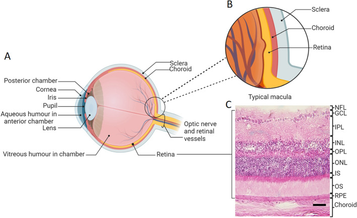
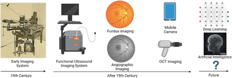
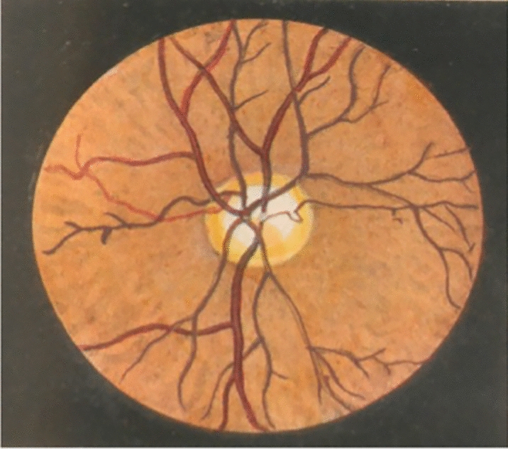
Recent years have seen significant advances in diagnostic testing of central nervous system (CNS) function and disease. However, there remain challenges in developing a comprehensive suite of non- or minimally invasive assays of neural health and disease progression. Due to the direct connection with the CNS, structural changes in the neural retina, retinal vasculature and morphological changes in retinal immune cells can occur in parallel with disease conditions in the brain. The retina can also, uniquely, be assessed directly and non-invasively. For these reasons, the retina may prove to be an important "window" for revealing and understanding brain disease. In this review, we discuss the gross anatomy of the eye, focusing on the sensory and non-sensory cells of the retina, especially microglia, that lend themselves to diagnosing brain disease by imaging the retina. We include a history of ocular imaging to describe the different imaging approaches undertaken in the past and outline current and emerging technologies including retinal autofluorescence imaging, Raman spectroscopy, and artificial intelligence image analysis. These new technologies show promising potential for retinal imaging to be used as a tool for the diagnosis of brain disorders such as Alzheimer's disease and others and the assessment of treatment success.
近年来,中枢神经系统(CNS)功能和疾病的诊断检测取得了重大进展。然而,在开发一套全面的神经健康和疾病进展的非侵入性或微创检测方法方面仍然存在挑战。由于与中枢神经系统直接相连,神经视网膜、视网膜血管和视网膜免疫细胞的形态变化可以与大脑疾病同时发生。视网膜还可以独特地直接进行非侵入性评估。出于这些原因,视网膜可能被证明是揭示和理解脑部疾病的一个重要“窗口”。在这篇综述中,我们讨论了眼睛的大体解剖结构,重点介绍了视网膜的感觉和非感觉细胞,特别是微胶质细胞,它们通过对视网膜进行成像来诊断脑部疾病。我们包括了眼部成像的历史,描述了过去所采用的不同成像方法,并概述了当前和新兴技术,包括视网膜自发荧光成像、拉曼光谱和人工智能图像分析。这些新技术显示出有希望的潜力,可将视网膜成像作为诊断阿尔茨海默病等脑部疾病以及评估治疗成功的工具。