文献检索文档翻译深度研究
Suppr Zotero 插件Zotero 插件
邀请有礼套餐&价格历史记录

新学期,新优惠

限时优惠:9月1日-9月22日

30天高级会员仅需29元

1天体验卡首发特惠仅需5.99元

了解详情
不再提醒
插件&应用
Suppr Zotero 插件Zotero 插件浏览器插件Mac 客户端Windows 客户端微信小程序
高级版
套餐订阅购买积分包
AI 工具
文献检索文档翻译深度研究
关于我们
关于 Suppr公司介绍联系我们用户协议隐私条款
关注我们

Suppr 超能文献

核心技术专利:CN118964589B侵权必究
粤ICP备2023148730 号-1Suppr @ 2025

尽管小胶质细胞被强烈激活,但光感受器丧失并不会募集中性粒细胞。

Photoreceptor loss does not recruit neutrophils despite strong microglial activation.

作者信息

Power Derek, Elstrott Justin, Schallek Jesse

机构信息

Center for Visual Science, University of Rochester, Rochester, United States.

Flaum Eye Institute, University of Rochester, Rochester, United States.

出版信息

Elife. 2025 Jul 22;13:RP98662. doi: 10.7554/eLife.98662.


DOI:10.7554/eLife.98662
PMID:40694413
原文链接:https://pmc.ncbi.nlm.nih.gov/articles/PMC12283074/
Abstract

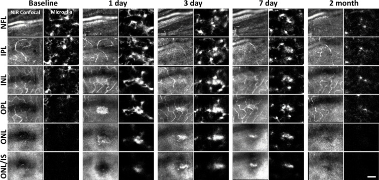
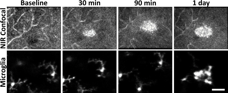
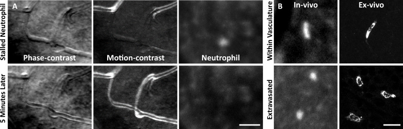
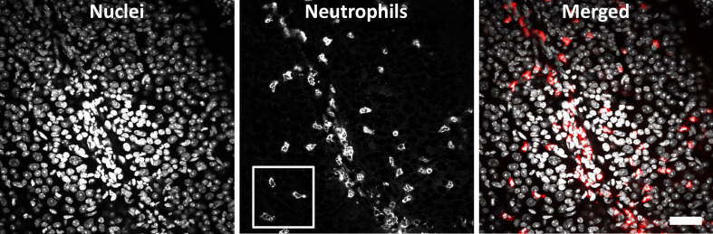
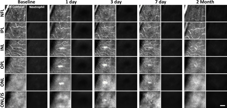
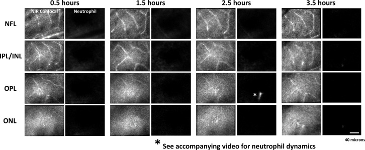
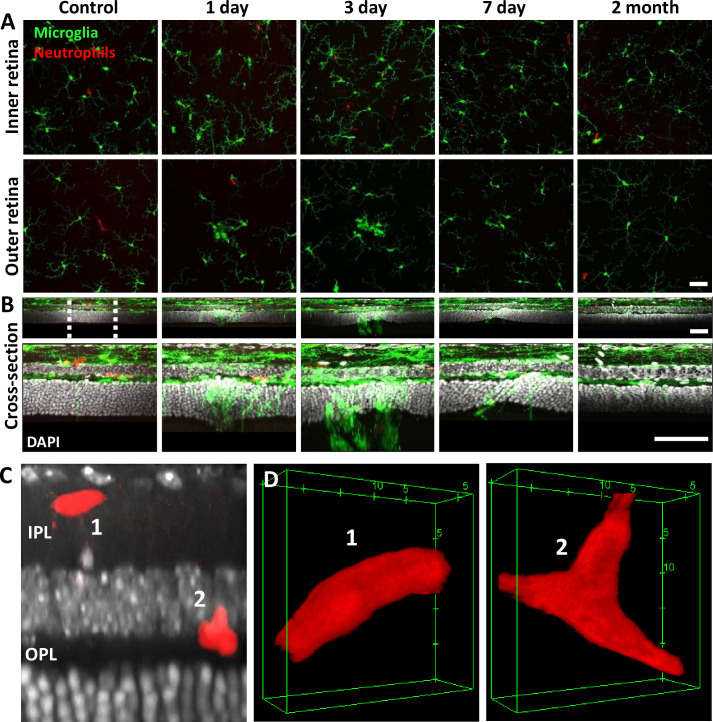
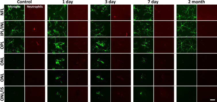
In response to central nervous system (CNS) injury, tissue-resident immune cells such as microglia and circulating systemic neutrophils are often first responders. The degree to which these cells interact in response to CNS damage is poorly understood, and even less so, in the neural retina, which poses a challenge for high-resolution imaging in vivo. In this study, we deploy fluorescence adaptive optics scanning light ophthalmoscopy (AOSLO) to study microglia and neutrophils in mice. We simultaneously track immune cell dynamics using label-free phase-contrast AOSLO at micron-level resolution. Retinal lesions were induced with 488 nm light focused onto photoreceptor (PR) outer segments. These lesions focally ablated PRs, with minimal collateral damage to cells above and below the plane of focus. We used in vivo AOSLO, and optical coherence tomography (OCT) imaging to reveal the natural history of the microglial and neutrophil response from minutes to months after injury. While microglia showed dynamic and progressive immune response with cells migrating into the injury locus within 1 day after injury, neutrophils were not recruited despite close proximity to vessels carrying neutrophils only microns away. Post-mortem confocal microscopy confirmed in vivo findings. This work illustrates that microglial activation does not recruit neutrophils in response to acute, focal loss of PRs, a condition encountered in many retinal diseases.

摘要

针对中枢神经系统(CNS)损伤,诸如小胶质细胞等组织驻留免疫细胞和循环系统中的中性粒细胞通常是首批响应者。人们对这些细胞在中枢神经系统损伤时相互作用的程度了解甚少,而在神经视网膜中更是知之甚少,这给体内高分辨率成像带来了挑战。在本研究中,我们采用荧光自适应光学扫描激光检眼镜(AOSLO)来研究小鼠中的小胶质细胞和中性粒细胞。我们使用无标记相衬AOSLO以微米级分辨率同时追踪免疫细胞动态。用聚焦于光感受器(PR)外段的488nm光诱导视网膜损伤。这些损伤局部消融PR,对焦点平面上方和下方的细胞造成的附带损伤最小。我们使用体内AOSLO和光学相干断层扫描(OCT)成像来揭示损伤后数分钟至数月内小胶质细胞和中性粒细胞反应的自然病程。虽然小胶质细胞表现出动态且渐进的免疫反应,细胞在损伤后1天内迁移至损伤部位,但尽管与仅几微米远的携带中性粒细胞的血管距离很近,中性粒细胞并未被募集。死后共聚焦显微镜检查证实了体内研究结果。这项工作表明,在许多视网膜疾病中出现的PR急性局灶性丧失的情况下,小胶质细胞激活不会募集中性粒细胞。

https://cdn.ncbi.nlm.nih.gov/pmc/blobs/71a2/12283074/abab050571fd/elife-98662-fig10.jpg
https://cdn.ncbi.nlm.nih.gov/pmc/blobs/71a2/12283074/6e95cb3e8c30/elife-98662-fig1.jpg
https://cdn.ncbi.nlm.nih.gov/pmc/blobs/71a2/12283074/5598a6aca4c2/elife-98662-fig1-figsupp1.jpg
https://cdn.ncbi.nlm.nih.gov/pmc/blobs/71a2/12283074/95f7ba45e052/elife-98662-fig2.jpg
https://cdn.ncbi.nlm.nih.gov/pmc/blobs/71a2/12283074/2e09fc82ba5d/elife-98662-fig3.jpg
https://cdn.ncbi.nlm.nih.gov/pmc/blobs/71a2/12283074/72ece5ae8253/elife-98662-fig4.jpg
https://cdn.ncbi.nlm.nih.gov/pmc/blobs/71a2/12283074/bb00d59f5f2e/elife-98662-fig4-figsupp1.jpg
https://cdn.ncbi.nlm.nih.gov/pmc/blobs/71a2/12283074/b234a7a8aba8/elife-98662-fig5.jpg
https://cdn.ncbi.nlm.nih.gov/pmc/blobs/71a2/12283074/cacb484171b9/elife-98662-fig6.jpg
https://cdn.ncbi.nlm.nih.gov/pmc/blobs/71a2/12283074/1ac4ca3908e0/elife-98662-fig6-figsupp1.jpg
https://cdn.ncbi.nlm.nih.gov/pmc/blobs/71a2/12283074/225924ed4ee7/elife-98662-fig7.jpg
https://cdn.ncbi.nlm.nih.gov/pmc/blobs/71a2/12283074/bd728bf62c5e/elife-98662-fig7-figsupp1.jpg
https://cdn.ncbi.nlm.nih.gov/pmc/blobs/71a2/12283074/ed6df07712ca/elife-98662-fig8.jpg
https://cdn.ncbi.nlm.nih.gov/pmc/blobs/71a2/12283074/27508abbdbc2/elife-98662-fig8-figsupp1.jpg
https://cdn.ncbi.nlm.nih.gov/pmc/blobs/71a2/12283074/c263a3b85b18/elife-98662-fig9.jpg
https://cdn.ncbi.nlm.nih.gov/pmc/blobs/71a2/12283074/f2a9f6ae6b30/elife-98662-fig9-figsupp1.jpg
https://cdn.ncbi.nlm.nih.gov/pmc/blobs/71a2/12283074/9ca0196f3385/elife-98662-fig9-figsupp2.jpg
https://cdn.ncbi.nlm.nih.gov/pmc/blobs/71a2/12283074/abab050571fd/elife-98662-fig10.jpg

相似文献

[1]
Photoreceptor loss does not recruit neutrophils despite strong microglial activation.

Elife. 2025-7-22

[2]
Photoreceptor loss does not recruit neutrophils despite strong microglial activation.

bioRxiv. 2025-3-25

[3]
Optical coherence tomography (OCT) for detection of macular oedema in patients with diabetic retinopathy.

Cochrane Database Syst Rev. 2015-1-7

[4]
Cone Density Changes After Repeated Low-Level Red Light Treatment in Children With Myopia.

JAMA Ophthalmol. 2025-4-24

[5]
Optic nerve head and fibre layer imaging for diagnosing glaucoma.

Cochrane Database Syst Rev. 2015-11-30

[6]
Optical coherence tomography (OCT) for detection of macular oedema in patients with diabetic retinopathy.

Cochrane Database Syst Rev. 2011-7-6

[7]
Interventions for central serous chorioretinopathy: a network meta-analysis.

Cochrane Database Syst Rev. 2025-6-16

[8]
Multimodal High-Resolution Imaging in Retinitis Pigmentosa: A Comparison Between Optoretinography, Cone Density, and Visual Sensitivity.

Invest Ophthalmol Vis Sci. 2024-8-1

[9]
Artificial intelligence for diagnosing exudative age-related macular degeneration.

Cochrane Database Syst Rev. 2024-10-17

[10]
Foveal Retinal Ganglion Cells Develop Altered Calcium Dynamics Weeks After Photoreceptor Ablation.

Ophthalmol Sci. 2024-3-22

引用本文的文献

[1]
Longitudinal High-Resolution Imaging of Retinal Sequelae of a Choroidal Nevus.

Diagnostics (Basel). 2025-7-29

本文引用的文献

[1]
Dysregulated CD200-CD200R signaling in early diabetes modulates microglia-mediated retinopathy.

Proc Natl Acad Sci U S A. 2023-11-7

[2]
High-resolution structural and functional retinal imaging in the awake behaving mouse.

Commun Biol. 2023-5-29

[3]
A laser-induced mouse model of progressive retinal degeneration with central sparing displays features of parafoveal geographic atrophy.

Sci Rep. 2023-3-14

[4]
Microglia-Neutrophil Interactions Drive Dry AMD-like Pathology in a Mouse Model.

Cells. 2022-11-9

[5]
Hyper-reflective retinal foci as possible in vivo imaging biomarker of microglia activation in von Hippel-Lindau disease.

PLoS One. 2022

[6]
Hyper-Reflecting Foci in Multiple Sclerosis Retina Associate With Macrophage/Microglia-Derived Cytokines in Cerebrospinal Fluid.

Front Immunol. 2022

[7]
In Vivo Capillary Structure and Blood Cell Flux in the Normal and Diabetic Mouse Eye.

Invest Ophthalmol Vis Sci. 2022-2-1

[8]
Imaging the dynamics of individual processes of microglia in the living retina .

Biomed Opt Express. 2021-9-10

[9]
Local photoreceptor cell death differences in the murine model of retinal detachment.

Sci Rep. 2021-9-22

[10]
The Neutrophil.

Immunity. 2021-7-13

文献AI研究员

20分钟写一篇综述,助力文献阅读效率提升50倍

立即体验

用中文搜PubMed

大模型驱动的PubMed中文搜索引擎

马上搜索

推荐工具

医学文档翻译智能文献检索