• 文献检索
  • 文档翻译
  • 深度研究
  • 学术资讯
  • Suppr Zotero 插件Zotero 插件
  • 邀请有礼
  • 套餐&价格
  • 历史记录
应用&插件
Suppr Zotero 插件Zotero 插件浏览器插件Mac 客户端Windows 客户端微信小程序
定价
高级版会员购买积分包购买API积分包
服务
文献检索文档翻译深度研究API 文档MCP 服务
关于我们
关于 Suppr公司介绍联系我们用户协议隐私条款
关注我们

Suppr 超能文献

核心技术专利:CN118964589B侵权必究
粤ICP备2023148730 号-1Suppr @ 2026

文献检索

告别复杂PubMed语法,用中文像聊天一样搜索,搜遍4000万医学文献。AI智能推荐,让科研检索更轻松。

立即免费搜索

文件翻译

保留排版,准确专业,支持PDF/Word/PPT等文件格式,支持 12+语言互译。

免费翻译文档

深度研究

AI帮你快速写综述,25分钟生成高质量综述,智能提取关键信息,辅助科研写作。

立即免费体验

2型黄斑毛细血管扩张症3至5期白内障手术后的视力

Visual acuity after cataract surgery in Macular Telangiectasia Type 2 Stage 3 to 5.

作者信息

Heng Jacob S, Arevalo J Fernando, Handa James T

机构信息

Department of Ophthalmology and Visual Science, Yale School of Medicine, New Haven, USA.

Department of Neuroscience, Johns Hopkins University School of Medicine, Baltimore, USA.

出版信息

Int J Retina Vitreous. 2022 Jun 11;8(1):38. doi: 10.1186/s40942-022-00386-0.

DOI:10.1186/s40942-022-00386-0
PMID:35690847
原文链接:https://pmc.ncbi.nlm.nih.gov/articles/PMC9188048/
Abstract

BACKGROUND

The purpose of this study was to evaluate visual acuity after cataract surgery in eyes with Macular Telangiectasia (MacTel) Type 2.

METHODS

Single-center retrospective cohort study of patients with MacTel Type 2 who underwent cataract surgery and were managed at the same institution. Patients underwent pre-operative assessment by a retinal specialist with examination and optical coherence tomography (OCT) at the same institution. The main outcome measure was the post-operative change in best corrected visual acuity (BCVA). Secondary study outcomes were achieving post-operative BCVA better than Snellen acuity of 20/40 and time to BCVA loss by two lines or more (10 or more ETDRS letters).

RESULTS

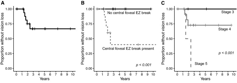
A total of 20 eyes (11 patients) underwent cataract surgery and were followed for a median of 25.5 months (IQR 17.5-44.2 months). The median post-operative BCVA improvement was 10.5 letters (IQR 3.50-20.25). Nuclear sclerosis severity [β = 8.99 (95% CI 3.35, 14.6), p = 0.00177] was associated with post-operative change in BCVA and central foveal ellipsoid zone (EZ) breaks [OR 1.33 × 10 (95% CI 5.12 × 10-3.43 × 10), p < 0.001] on OCT was inversely correlated with post-operative BCVA > 20/40 using a multivariate generalized linear model. Central foveal EZ breaks [HR 1.77 × 10 (95% CI 3.86 × 10, 8.11 × 10), p < 0.001] and MacTel Type 2 disease stage [HR 2.83, (95% CI 1.12, 7.12), p = 0.027] were independently associated with shorter time to vision loss of two lines or more in a multivariate Cox regression model.

CONCLUSIONS

Visual acuity significant improved after cataract surgery in eyes with MacTel Type 2 regardless of disease severity. The presence of central foveal EZ breaks may predict poorer post-operative visual acuity and subsequent vision loss from disease progression.

摘要

背景

本研究的目的是评估2型黄斑毛细血管扩张症(MacTel)患者白内障手术后的视力。

方法

对在同一机构接受白内障手术并接受治疗的2型MacTel患者进行单中心回顾性队列研究。患者在同一机构由视网膜专科医生进行术前评估,包括检查和光学相干断层扫描(OCT)。主要结局指标是最佳矫正视力(BCVA)的术后变化。次要研究结局是术后BCVA优于Snellen视力20/40,以及BCVA下降两行或更多(10个或更多ETDRS字母)的时间。

结果

共有20只眼(11例患者)接受了白内障手术,中位随访时间为25.5个月(四分位间距17.5 - 44.2个月)。术后BCVA改善的中位数为10.5个字母(四分位间距3.50 - 20.25)。核硬化严重程度[β = 8.99(95%可信区间3.35, 14.6),p = 0.00177]与BCVA的术后变化相关,并且在多变量广义线性模型中,OCT上的中央凹椭圆体带(EZ)断裂[比值比1.33×10(95%可信区间5.12×10 - 3.43×10),p < 0.001]与术后BCVA > 20/40呈负相关。在多变量Cox回归模型中,中央凹EZ断裂[风险比1.77×10(95%可信区间3.86×10, 8.11×10),p < 0.001]和2型MacTel疾病分期[风险比2.83,(95%可信区间1.12, 7.12),p = 0.027]与视力下降两行或更多的时间缩短独立相关。

结论

无论疾病严重程度如何,2型MacTel患者白内障手术后视力均有显著改善。中央凹EZ断裂的存在可能预示术后视力较差以及疾病进展导致的后续视力丧失。

https://cdn.ncbi.nlm.nih.gov/pmc/blobs/c3c9/9188048/588c9fe20b38/40942_2022_386_Fig2_HTML.jpg
https://cdn.ncbi.nlm.nih.gov/pmc/blobs/c3c9/9188048/768e0c102a51/40942_2022_386_Fig1_HTML.jpg
https://cdn.ncbi.nlm.nih.gov/pmc/blobs/c3c9/9188048/588c9fe20b38/40942_2022_386_Fig2_HTML.jpg
https://cdn.ncbi.nlm.nih.gov/pmc/blobs/c3c9/9188048/768e0c102a51/40942_2022_386_Fig1_HTML.jpg
https://cdn.ncbi.nlm.nih.gov/pmc/blobs/c3c9/9188048/588c9fe20b38/40942_2022_386_Fig2_HTML.jpg

相似文献

1
Visual acuity after cataract surgery in Macular Telangiectasia Type 2 Stage 3 to 5.2型黄斑毛细血管扩张症3至5期白内障手术后的视力
Int J Retina Vitreous. 2022 Jun 11;8(1):38. doi: 10.1186/s40942-022-00386-0.
2
Long-term Outcomes of Cataract Surgery in Patients with Retinitis Pigmentosa.色素性视网膜炎患者白内障手术的长期疗效。
Ophthalmol Retina. 2022 Apr;6(4):268-272. doi: 10.1016/j.oret.2021.12.010. Epub 2021 Dec 16.
3
Macular Telangiectasia Type 2: Visual Acuity, Disease End Stage, and the MacTel Area: MacTel Project Report Number 8.特发性脉络膜新生血管(湿性年龄相关性黄斑变性)2 型:视力、疾病终末期和 MacTel 区:MacTel 项目报告第 8 号。
Ophthalmology. 2020 Nov;127(11):1539-1548. doi: 10.1016/j.ophtha.2020.03.040. Epub 2020 Apr 21.
4
CORRELATION OF CLINICAL AND STRUCTURAL PROGRESSION WITH VISUAL ACUITY LOSS IN MACULAR TELANGIECTASIA TYPE 2: MacTel Project Report No. 6-The MacTel Research Group.2型黄斑毛细血管扩张症临床及结构进展与视力丧失的相关性:黄斑毛细血管扩张症项目报告第6号——黄斑毛细血管扩张症研究组
Retina. 2018 Jan;38 Suppl 1(Suppl 1):S8-S13. doi: 10.1097/IAE.0000000000001697.
5
Macular Telangiectasia Type 2: A Classification System Using MultiModal Imaging MacTel Project Report Number 10.2型黄斑毛细血管扩张症:一种使用多模态成像的分类系统。MacTel项目报告编号10。
Ophthalmol Sci. 2022 Dec 8;3(2):100261. doi: 10.1016/j.xops.2022.100261. eCollection 2023 Jun.
6
Progression of vascular changes in macular telangiectasia type 2: comparison between SD-OCT and OCT angiography.2型黄斑毛细血管扩张症血管变化的进展:频域光学相干断层扫描与光学相干断层扫描血管造影的比较
Graefes Arch Clin Exp Ophthalmol. 2019 Jul;257(7):1381-1392. doi: 10.1007/s00417-019-04323-0. Epub 2019 May 16.
7
Visual Acuity in Retinal Vein Occlusion, Diabetic, and Uveitic Macular Edema: Central Subfield Thickness and Ellipsoid Zone Analysis.视网膜静脉阻塞、糖尿病性和葡萄膜炎性黄斑水肿的视力:中心凹下视网膜厚度和椭圆体带分析。
Ophthalmol Retina. 2021 Jul;5(7):633-647. doi: 10.1016/j.oret.2020.10.016. Epub 2020 Oct 29.
8
OCT Angiography and Ellipsoid Zone Mapping of Macular Telangiectasia Type 2 From the AVATAR Study.来自AVATAR研究的2型黄斑毛细血管扩张症的光学相干断层扫描血管造影及椭圆体带成像
Invest Ophthalmol Vis Sci. 2017 Jul 1;58(9):3683-3689. doi: 10.1167/iovs.16-20976.
9
Retinal cavitations in macular telangiectasia type 2 (MacTel): longitudinal structure-function correlations.视网膜囊腔在 2 型黄斑毛细血管扩张症(MacTel)中的表现:纵向结构-功能相关性。
Br J Ophthalmol. 2021 Jan;105(1):109-112. doi: 10.1136/bjophthalmol-2019-315416. Epub 2020 Mar 9.
10
Progression characteristics of ellipsoid zone loss in macular telangiectasia type 2.椭圆体带丢失在 2 型黄斑毛细血管扩张症中的进展特征。
Acta Ophthalmol. 2019 Nov;97(7):e998-e1005. doi: 10.1111/aos.14110. Epub 2019 Apr 9.

本文引用的文献

1
Retinal cavitations in macular telangiectasia type 2 (MacTel): longitudinal structure-function correlations.视网膜囊腔在 2 型黄斑毛细血管扩张症(MacTel)中的表现:纵向结构-功能相关性。
Br J Ophthalmol. 2021 Jan;105(1):109-112. doi: 10.1136/bjophthalmol-2019-315416. Epub 2020 Mar 9.
2
Hypoxia tolerance in the Norrin-deficient retina and the chronically hypoxic brain studied at single-cell resolution.在单细胞分辨率水平上研究 Norrin 缺陷型视网膜和慢性缺氧大脑的耐缺氧能力。
Proc Natl Acad Sci U S A. 2019 Apr 30;116(18):9103-9114. doi: 10.1073/pnas.1821122116. Epub 2019 Apr 15.
3
Progression characteristics of ellipsoid zone loss in macular telangiectasia type 2.
椭圆体带丢失在 2 型黄斑毛细血管扩张症中的进展特征。
Acta Ophthalmol. 2019 Nov;97(7):e998-e1005. doi: 10.1111/aos.14110. Epub 2019 Apr 9.
4
ELECTROPHYSIOLOGICAL CHARACTERIZATION OF MACULAR TELANGIECTASIA TYPE 2 AND STRUCTURE-FUNCTION CORRELATION.2型黄斑毛细血管扩张症的电生理特征及结构-功能相关性
Retina. 2018 Jan;38 Suppl 1:S33-S42. doi: 10.1097/IAE.0000000000001746.
5
LONGITUDINAL CORRELATION OF ELLIPSOID ZONE LOSS AND FUNCTIONAL LOSS IN MACULAR TELANGIECTASIA TYPE 2.2型黄斑毛细血管扩张症中椭球体带丢失与功能丧失的纵向相关性
Retina. 2018 Jan;38 Suppl 1(Suppl 1):S20-S26. doi: 10.1097/IAE.0000000000001715.
6
CORRELATION OF CLINICAL AND STRUCTURAL PROGRESSION WITH VISUAL ACUITY LOSS IN MACULAR TELANGIECTASIA TYPE 2: MacTel Project Report No. 6-The MacTel Research Group.2型黄斑毛细血管扩张症临床及结构进展与视力丧失的相关性:黄斑毛细血管扩张症项目报告第6号——黄斑毛细血管扩张症研究组
Retina. 2018 Jan;38 Suppl 1(Suppl 1):S8-S13. doi: 10.1097/IAE.0000000000001697.
7
Genome-wide analyses identify common variants associated with macular telangiectasia type 2.全基因组分析鉴定出与黄斑毛细血管扩张症 2 型相关的常见变异。
Nat Genet. 2017 Apr;49(4):559-567. doi: 10.1038/ng.3799. Epub 2017 Feb 27.
8
Progression of Vision Loss in Macular Telangiectasia Type 2.2型黄斑毛细血管扩张症视力丧失的进展
Invest Ophthalmol Vis Sci. 2015 Jun;56(6):3905-12. doi: 10.1167/iovs.15-16915.
9
Loss of Müller's cells and photoreceptors in macular telangiectasia type 2.Müller 细胞和光感受器在 2 型黄斑毛细血管扩张症中的丧失。
Ophthalmology. 2013 Nov;120(11):2344-52. doi: 10.1016/j.ophtha.2013.04.013. Epub 2013 Jun 12.
10
Macular telangiectasia type 2.2 型黄斑毛细血管扩张症。
Prog Retin Eye Res. 2013 May;34:49-77. doi: 10.1016/j.preteyeres.2012.11.002. Epub 2012 Dec 3.