Liu Yinghong, He Mingyue, Xiong Hao, Yuan Fang
Hunan Key Laboratory of Kidney Disease and Blood Purification, Department of Nephrology, The Second Xiangya Hospital of Central South University, Changsha, China.
Front Med (Lausanne). 2022 May 27;9:874916. doi: 10.3389/fmed.2022.874916. eCollection 2022.
The micro-inflammatory state is important for the occurrence of diabetic kidney disease (DKD). Here, we aimed to explore the expression of pyroptosis related indicators and ultrastructural characteristics in DKD, and investigate pyroptosis in renal tubular epithelial cells induced by high glucose.
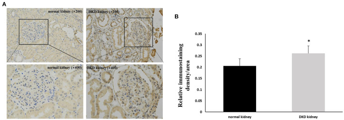
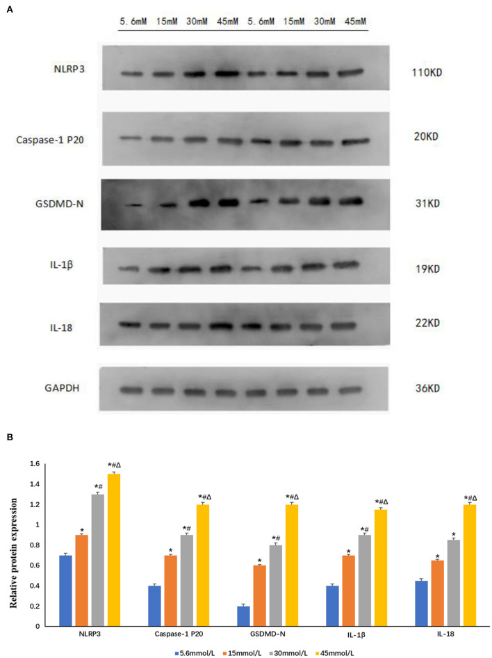
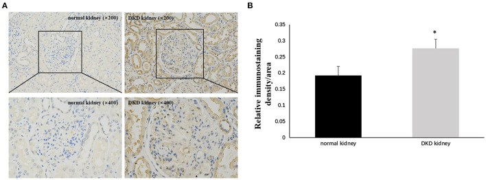
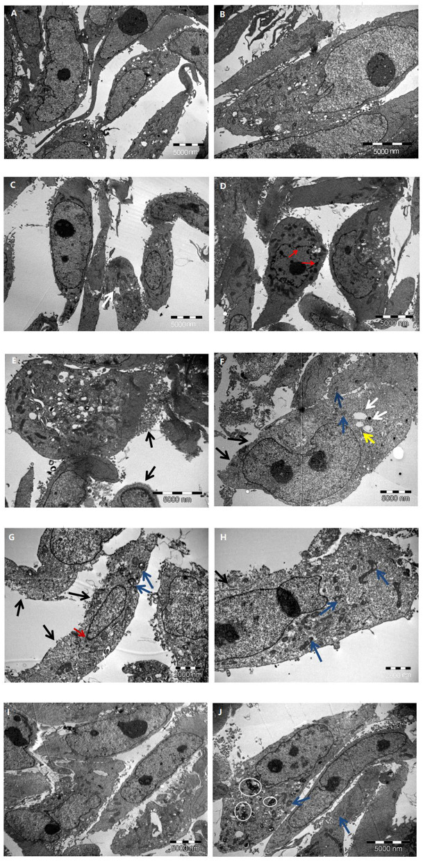
Immunohistochemistry was used to detect expression of the inflammation-related protein NOD-like receptor protein 3 (NLRP3) and pyroptosis key protein gasdermin D (GSDMD) in kidney tissues of DKD patients. HK-2 cells were cultured and stimulated with different concentrations of glucose. The changes in HK-2 cell ultrastructure were observed using electronmicroscopy, and western blot was used to detect NLRP3, caspase-1 p20, GSDMD-N, interleukin (IL)-1β, and IL-18 expression.
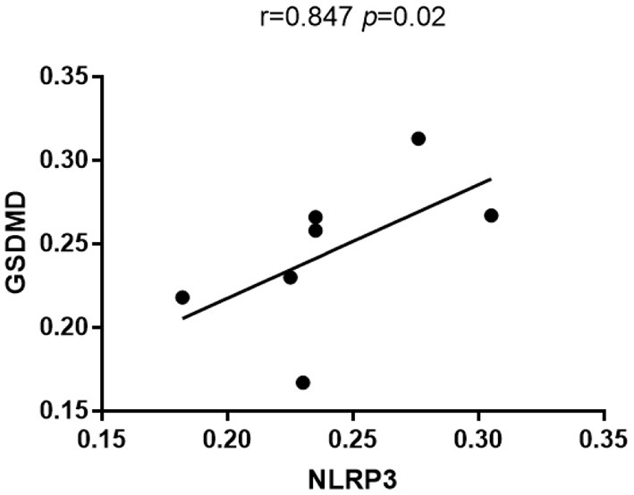
NLRP3 and GSDMD expression in kidney tissues of DKD patients was higher than that in control subjects. Further, GSDMD expression was positively correlated with that of NLRP3 ( = 0.847, = 0.02). After stimulating HK-2 cells for 24 h with different glucose concentrations, compared with the control group, the 15 and 30 mmol/L glucose groups showed typical ultrastructural changes of pyroptosis. The protein expression of NLRP3, caspase-1 p20, GSDMD-N, IL-1β, and IL-18 expression in high glucose group increased significantly compared with the control group, and was glucose-concentration-dependent.
High glucose can activate inflammasome, cause inflammatory cytokines release, and induce pyroptosis in HK-2 cells. NLRP3-caspase-1 may be involved in GSDMD-mediated pyroptosis. This study shows a novel relationship between glucose concentration and pyroptosis, which can be studied further to design better therapies for patients with DKD.
微炎症状态对糖尿病肾病(DKD)的发生发展至关重要。在此,我们旨在探究DKD中细胞焦亡相关指标的表达及超微结构特征,并研究高糖诱导肾小管上皮细胞发生细胞焦亡的情况。
采用免疫组化法检测DKD患者肾组织中炎症相关蛋白NOD样受体蛋白3(NLRP3)和细胞焦亡关键蛋白gasdermin D(GSDMD)的表达。培养HK-2细胞并用不同浓度葡萄糖进行刺激。利用电子显微镜观察HK-2细胞超微结构的变化,采用蛋白质印迹法检测NLRP3、半胱天冬酶-1 p20、GSDMD-N、白细胞介素(IL)-1β和IL-18的表达。
DKD患者肾组织中NLRP3和GSDMD的表达高于对照组。此外,GSDMD的表达与NLRP3的表达呈正相关(r = 0.847,P = 0.02)。用不同葡萄糖浓度刺激HK-2细胞24小时后,与对照组相比,15 mmol/L和30 mmol/L葡萄糖组出现了典型的细胞焦亡超微结构变化。高糖组中NLRP3、半胱天冬酶-1 p20、GSDMD-N、IL-1β和IL-18的蛋白表达与对照组相比显著增加,且呈葡萄糖浓度依赖性。
高糖可激活炎性小体,导致炎性细胞因子释放,并诱导HK-2细胞发生细胞焦亡。NLRP3-半胱天冬酶-1可能参与GSDMD介导的细胞焦亡。本研究揭示了葡萄糖浓度与细胞焦亡之间的新关系,可进一步深入研究,为DKD患者设计更好的治疗方案。