• 文献检索
  • 文档翻译
  • 深度研究
  • 学术资讯
  • Suppr Zotero 插件Zotero 插件
  • 邀请有礼
  • 套餐&价格
  • 历史记录
应用&插件
Suppr Zotero 插件Zotero 插件浏览器插件Mac 客户端Windows 客户端微信小程序
定价
高级版会员购买积分包购买API积分包
服务
文献检索文档翻译深度研究API 文档MCP 服务
关于我们
关于 Suppr公司介绍联系我们用户协议隐私条款
关注我们

Suppr 超能文献

核心技术专利:CN118964589B侵权必究
粤ICP备2023148730 号-1Suppr @ 2026

文献检索

告别复杂PubMed语法,用中文像聊天一样搜索,搜遍4000万医学文献。AI智能推荐,让科研检索更轻松。

立即免费搜索

文件翻译

保留排版,准确专业,支持PDF/Word/PPT等文件格式,支持 12+语言互译。

免费翻译文档

深度研究

AI帮你快速写综述,25分钟生成高质量综述,智能提取关键信息,辅助科研写作。

立即免费体验

联动成像在经鼻内镜检查中能有效检测内镜下萎缩边界。

Linked color imaging effectively detects the endoscopic atrophic border in transnasal endoscopy.

作者信息

Kawai Yusuke, Sugimoto Mitsushige, Hamada Mariko, Iwata Eri, Niikura Ryota, Nagata Naoyoshi, Fukuzawa Masakatsu, Itoi Takao, Kawai Takashi

机构信息

Department of Gastroenterological Endoscopy, Tokyo Medical University Hospital, 6-7-1 Nishishinjuku, Shinjuku-ku, Tokyo 160-0023, Japan.

Department of Gastroenterology, Tokyo Medical University Hospital, 6-7-1 Nishishinjuku, Shinjuku-ku, Tokyo 160-0023, Japan.

出版信息

J Clin Biochem Nutr. 2022 May;70(3):290-296. doi: 10.3164/jcbn.21-145. Epub 2022 Feb 1.

DOI:10.3164/jcbn.21-145
PMID:35692679
原文链接:https://pmc.ncbi.nlm.nih.gov/articles/PMC9130059/
Abstract

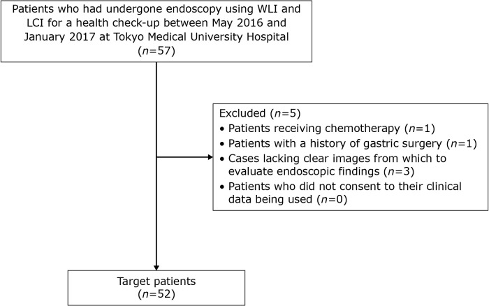
In oral endoscopy, linked color imaging (LCI) detects atrophic border and gastric mucosal diseases better than white light imaging (WLI), but its usefulness in transnasal endoscopy has not been fully investigated. Here, we retrospectively compared WLI and LCI using the Lab* color space in images from 57 patients aged ≥20 years who had undergone transnasal endoscopy as part of a health check-up from May 2016 to January 2017. We measured color differences at the atrophic/non-atrophic and fundic/pyloric mucosal borders. Gastritis severity scored using the Kyoto classification of gastritis was similar between the two techniques. However, in patients with current and with past infection, color difference at the atrophic border was greater with LCI (21.58 ± 6.97 and 27.34 ± 10.32, respectively) than with WLI [14.42 ± 5.95 ( = 0.004) and 17.9 ± 8.48 (<0.001)]; in those never infected with , color difference at the fundic/pyloric mucosal border was greater with LCI than with WLI (<0.001). Because of its enhancement of atrophic border detection, we recommend linked color imaging as the method of choice for transnasal endoscopy in health check-ups, particularly for identifying people at high risk of gastric cancer.

摘要

在口腔内镜检查中,与白光成像(WLI)相比,联动成像(LCI)能更好地检测萎缩边界和胃黏膜疾病,但它在鼻内镜检查中的应用价值尚未得到充分研究。在此,我们回顾性比较了2016年5月至2017年1月期间作为健康检查一部分接受鼻内镜检查的57例年龄≥20岁患者图像中使用Lab*颜色空间的WLI和LCI。我们测量了萎缩/非萎缩和胃底/幽门黏膜边界处的颜色差异。两种技术使用胃炎京都分类法评分的胃炎严重程度相似。然而,在现感染和既往感染患者中,LCI在萎缩边界处的颜色差异(分别为21.58±6.97和27.34±10.32)大于WLI[14.42±5.95(P = 0.004)和17.9±8.48(P<0.001)];在从未感染过幽门螺杆菌的患者中,LCI在胃底/幽门黏膜边界处的颜色差异大于WLI(P<0.001)。由于其增强了萎缩边界检测,我们建议在健康检查中联动成像作为鼻内镜检查的首选方法,特别是用于识别胃癌高危人群。

https://cdn.ncbi.nlm.nih.gov/pmc/blobs/d1ea/9130059/cc53bbb874a4/jcbn21-145f01.jpg

相似文献

1
Linked color imaging effectively detects the endoscopic atrophic border in transnasal endoscopy.联动成像在经鼻内镜检查中能有效检测内镜下萎缩边界。
J Clin Biochem Nutr. 2022 May;70(3):290-296. doi: 10.3164/jcbn.21-145. Epub 2022 Feb 1.
2
Objective Endoscopic Analysis with Linked Color Imaging regarding Gastric Mucosal Atrophy: A Pilot Study.关于胃黏膜萎缩的内镜下联合彩色成像分析:一项初步研究。
Gastroenterol Res Pract. 2017;2017:5054237. doi: 10.1155/2017/5054237. Epub 2017 Nov 15.
3
Efficacy of high-vision transnasal endoscopy using texture and colour enhancement imaging and narrow-band imaging to evaluate gastritis: a randomized controlled trial.高分辨率经鼻内镜联合纹理增强及窄带成像技术评估胃炎的疗效:一项随机对照试验。
Ann Med. 2022 Dec;54(1):1004-1013. doi: 10.1080/07853890.2022.2063372.
4
Enhanced Visibility in Evaluating Gastric Cancer and Helicobacter pylori-Associated Gastritis Using Linked Color Imaging with a Light-Emitting Diode Light Source.使用发光二极管光源的蓝激光成像增强对胃癌和幽门螺杆菌相关性胃炎的可视性。
Dig Dis Sci. 2022 Jun;67(6):2367-2374. doi: 10.1007/s10620-021-07234-5. Epub 2021 Aug 31.
5
The role of linked color imaging in endoscopic diagnosis of associated gastritis.链接色成像在相关胃炎内镜诊断中的作用。
Scand J Gastroenterol. 2020 Sep;55(9):1114-1120. doi: 10.1080/00365521.2020.1794025. Epub 2020 Jul 15.
6
Accuracies of Endoscopic Diagnosis of Helicobacter pylori-Gastritis: Multicenter Prospective Study Using White Light Imaging and Linked Color Imaging.幽门螺杆菌胃炎的内镜诊断准确性:使用白光成像和链接彩色成像的多中心前瞻性研究。
Digestion. 2020;101(5):624-630. doi: 10.1159/000501634. Epub 2019 Jul 23.
7
Linked Color Imaging and the Kyoto Classification of Gastritis: Evaluation of Visibility and Inter-Rater Reliability.关联显色内镜与京都胃炎分类:可视度评估与观察者间可靠性研究。
Digestion. 2020;101(5):598-607. doi: 10.1159/000501534. Epub 2019 Jul 12.
8
Detection of early stage gastric cancers in screening laser endoscopy using linked color imaging for patients with atrophic gastritis.应用连接色彩成像技术对萎缩性胃炎患者进行筛查性激光内视镜检查以早期发现胃癌。
J Gastroenterol Hepatol. 2021 Jun;36(6):1642-1648. doi: 10.1111/jgh.15312. Epub 2020 Nov 10.
9
Linked color imaging improves endoscopic diagnosis of active Helicobacter pylori infection.联动成像可提高内镜对活动性幽门螺杆菌感染的诊断能力。
Endosc Int Open. 2016 Jul;4(7):E800-5. doi: 10.1055/s-0042-109049.
10
A prospective randomized tandem gastroscopy pilot study of linked color imaging versus white light imaging for detection of upper gastrointestinal lesions.一项关于联合彩色成像与白光成像检测上消化道病变的前瞻性随机串联胃镜试点研究。
J Gastroenterol Hepatol. 2021 Sep;36(9):2562-2567. doi: 10.1111/jgh.15515. Epub 2021 Apr 12.

引用本文的文献

1
Unusual Cause of Duodenal Villi Atrophy.十二指肠绒毛萎缩的罕见病因。
Dig Dis Sci. 2023 Oct;68(10):3827-3828. doi: 10.1007/s10620-023-08074-1. Epub 2023 Aug 21.
2
Linked Color Imaging for Stomach.胃部的联动彩色成像
Diagnostics (Basel). 2023 Jan 27;13(3):467. doi: 10.3390/diagnostics13030467.
3
Visibility of early gastric cancers by texture and color enhancement imaging using a high-definition ultrathin transnasal endoscope.使用高清超微经鼻内窥镜的纹理和颜色增强成像技术对早期胃癌的可视性。

本文引用的文献

1
AGA Clinical Practice Update on the Diagnosis and Management of Atrophic Gastritis: Expert Review.美国胃肠病学会关于萎缩性胃炎诊断与管理的临床实践更新:专家综述
Gastroenterology. 2021 Oct;161(4):1325-1332.e7. doi: 10.1053/j.gastro.2021.06.078. Epub 2021 Aug 26.
2
Gastric mucosal changes, and sex differences therein, after Helicobacter pylori eradication: A long-term prospective follow-up study.幽门螺杆菌根除后胃黏膜的变化及其性别差异:一项长期前瞻性随访研究。
J Gastroenterol Hepatol. 2021 Aug;36(8):2210-2216. doi: 10.1111/jgh.15477. Epub 2021 Mar 12.
3
The simplified Kyoto classification score is consistent with the ABC method of classification as a grading system for endoscopic gastritis.
Sci Rep. 2023 Feb 3;13(1):1994. doi: 10.1038/s41598-023-29284-7.
4
Using texture and colour enhancement imaging to evaluate gastrointestinal diseases in clinical practice: a review.利用纹理和色彩增强成像技术评估临床胃肠道疾病:综述。
Ann Med. 2022 Dec;54(1):3315-3332. doi: 10.1080/07853890.2022.2147992.
5
Efficacy of a Third-Generation High-Vision Ultrathin Endoscope for Evaluating Gastric Atrophy and Intestinal Metaplasia in -Eradicated Patients.第三代高清超薄内镜在评估根除幽门螺杆菌患者胃萎缩和肠化生中的疗效
J Clin Med. 2022 Apr 14;11(8):2198. doi: 10.3390/jcm11082198.
简化的京都分类评分与作为内镜下胃炎分级系统的ABC分类方法一致。
J Clin Biochem Nutr. 2021 Jan;68(1):101-104. doi: 10.3164/jcbn.20-41. Epub 2020 Jul 16.
4
Linked Color Imaging Focused on Neoplasm Detection in the Upper Gastrointestinal Tract : A Randomized Trial.链接染色成像在上消化道肿瘤检测中的应用:一项随机试验。
Ann Intern Med. 2021 Jan;174(1):18-24. doi: 10.7326/M19-2561. Epub 2020 Oct 20.
5
Optical effect of spraying l-menthol on gastric intestinal metaplasia visualized by linked color imaging.喷雾薄荷醇对胃肠化生的可视光学效应。
Eur J Gastroenterol Hepatol. 2021 Mar 1;33(3):358-363. doi: 10.1097/MEG.0000000000001910.
6
Chemoprevention of gastric cancer development after eradication therapy in an East Asian population: Meta-analysis.东亚人群根除治疗后胃癌发展的化学预防:荟萃分析。
World J Gastroenterol. 2020 Apr 21;26(15):1820-1840. doi: 10.3748/wjg.v26.i15.1820.
7
An evidence-based guideline on the application of molecular testing in the diagnosis, prediction of prognosis, and selection of therapy in non-GIST soft tissue sarcomas.非 GIST 软组织肉瘤分子检测在诊断、预后预测和治疗选择中的应用的循证指南。
Cancer Treat Rev. 2020 Apr;85:101987. doi: 10.1016/j.ctrv.2020.101987. Epub 2020 Feb 14.
8
Accuracies of Endoscopic Diagnosis of Helicobacter pylori-Gastritis: Multicenter Prospective Study Using White Light Imaging and Linked Color Imaging.幽门螺杆菌胃炎的内镜诊断准确性:使用白光成像和链接彩色成像的多中心前瞻性研究。
Digestion. 2020;101(5):624-630. doi: 10.1159/000501634. Epub 2019 Jul 23.
9
Validity of endoscopic features for the diagnosis of Helicobacter pylori infection status based on the Kyoto classification of gastritis.基于胃炎京都分类的内镜特征对幽门螺杆菌感染状态的诊断价值。
Dig Endosc. 2020 Jan;32(1):74-83. doi: 10.1111/den.13486. Epub 2019 Sep 26.
10
Linked Color Imaging and the Kyoto Classification of Gastritis: Evaluation of Visibility and Inter-Rater Reliability.关联显色内镜与京都胃炎分类:可视度评估与观察者间可靠性研究。
Digestion. 2020;101(5):598-607. doi: 10.1159/000501534. Epub 2019 Jul 12.