Issashar Leibovitzh Galia, Trope Graham E, Buys Yvonne M, Tarita-Nistor Luminita
Krembil Research Institute, Donald K. Johnson Eye Institute, University Health Network, Toronto, ON, Canada.
Department of Ophthalmology and Vision Sciences, Toronto Western Hospital, Toronto, ON, Canada.
Front Aging Neurosci. 2022 May 25;14:833150. doi: 10.3389/fnagi.2022.833150. eCollection 2022.
This study tested perceptual grouping during binocular rivalry to probe the strength of neural connectivity of the visual cortex involved in early visual processing in patients with mild glaucoma.
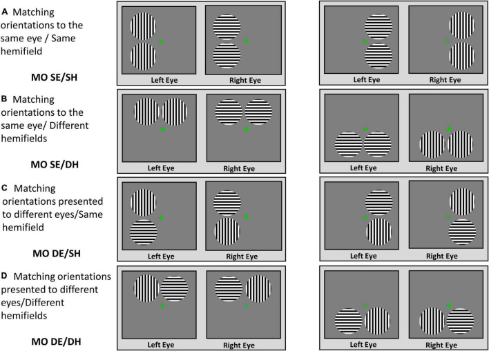
Seventeen patients with mild glaucoma with no significant visual field defects and 14 healthy controls participated. Rivalry stimuli were 1.8°-diameter discs, containing horizontal or vertical sine-wave gratings, viewed dichoptically. To test the grouping, two spatially separated identical stimuli were presented eccentrically to the same or different eyes and to the same or different hemifields. The outcome measures were the time of exclusive dominance of the grouped percept (i.e., percept with synchronized orientations), the rivalry rate, and the epochs of exclusive dominance.
For both groups, the grouping occurred primarily for the matching orientations in the same eye/same hemifield (MO SE/SH) and for the matching orientations in the same eye/different hemifield (MO SE/DH) conditions. Time dominance of the grouped percept of the glaucoma group was similar to that of the control group in all conditions. The rivalry rates in the MO SE/SH and MO SE/DH conditions were significantly larger in the control group than in the glaucoma group. The epochs of exclusive dominance of the grouped percept in the MO SE/SH condition were a median of 48-ms longer for the control group, but a median of 116-ms shorter for the glaucoma group when compared to those in the MO SE/DH condition.
Patients with mild glaucoma show clear impairments in binocular rivalry while evidence for deficits in perceptual grouping could be inferred only indirectly. If these deficits truly exist, they may have implications for higher levels of visual processing, such as object recognition and scene segmentation, but these predictions remain to be tested in future studies.
本研究通过双眼竞争期间的知觉分组来探究轻度青光眼患者早期视觉处理中涉及的视觉皮层神经连接强度。
17例无明显视野缺损的轻度青光眼患者和14名健康对照者参与研究。竞争刺激为直径1.8°的圆盘,包含水平或垂直正弦波光栅,采用双眼分视呈现。为测试分组情况,将两个空间分离的相同刺激偏心地呈现给同一只眼或不同眼以及同侧或异侧半视野。结果测量指标为分组知觉(即方向同步的知觉)的独占主导时间、竞争率和独占主导期。
对于两组,分组主要发生在同一只眼/同侧半视野(MO SE/SH)的匹配方向以及同一只眼/异侧半视野(MO SE/DH)的匹配方向条件下。青光眼组在所有条件下分组知觉的时间主导与对照组相似。对照组在MO SE/SH和MO SE/DH条件下的竞争率显著高于青光眼组。与MO SE/DH条件相比,对照组在MO SE/SH条件下分组知觉的独占主导期中位数长48毫秒,而青光眼组则短116毫秒。
轻度青光眼患者在双眼竞争中表现出明显受损,而仅能间接推断出知觉分组存在缺陷的证据。如果这些缺陷确实存在,可能会对更高层次的视觉处理产生影响,如物体识别和场景分割,但这些预测仍有待未来研究进行验证。