Department of Ophthalmology, School of Medicine, Kitasato University, Kanagawa, Japan.
Moorfields Eye Hospital NHS Foundation Trust and University College London, Institute of Ophthalmology, London, United Kingdom.
PLoS One. 2018 Nov 21;13(11):e0207517. doi: 10.1371/journal.pone.0207517. eCollection 2018.
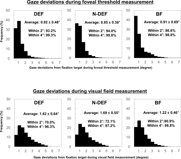
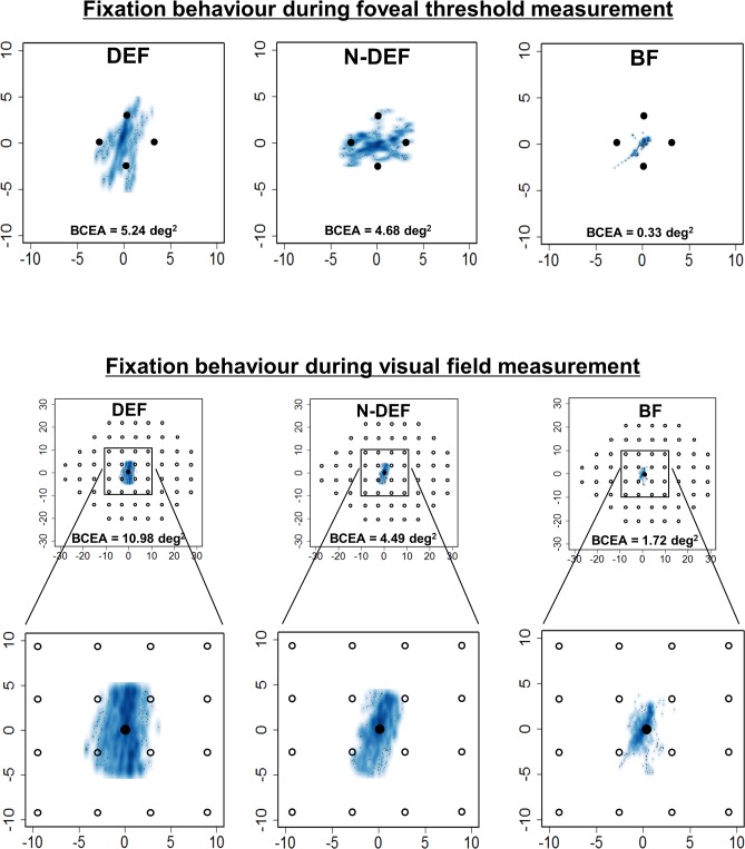
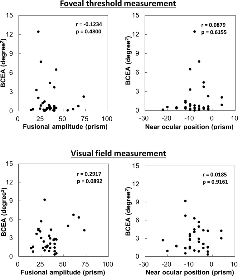
The aim of this cross-sectional study was to use standard automated perimetry to compare fixation variability among the dominant eye fixation, non-dominant eye fixation, and binocular fixation conditions. Thirty-five eyes of 35 healthy young participants underwent standard automated perimetry (Humphrey 24-2 SITA-Standard) in dominant eye fixation, non-dominant eye fixation, and binocular fixation conditions. Fixation variability during foveal threshold and visual field measurement, which was recorded using a wearable eye-tracking glass and calculated using the bivariate contour ellipse area (deg2), was compared among the three fixation conditions. Further, the association of bivariate contour ellipse area with ocular position and fusional amplitude during binocular fixation was analysed. There were no significant differences in bivariate contour ellipse area during foveal threshold measurement among the dominant eye fixation (1.75 deg2), non-dominant eye fixation (1.45 deg2), and binocular fixation (1.62 deg2) conditions. In contrast, the bivariate contour ellipse area during visual field measurement in binocular fixation (2.85 deg2) was significantly lower than the bivariate contour ellipse area in dominant eye fixation (4.62 deg2; p = 0.0227) and non-dominant eye fixation (5.24 deg2; p = 0.0006) conditions. There was no significant difference in bivariate contour ellipse area during visual field measurement between dominant eye fixation and non-dominant eye fixation conditions. There was no significant correlation between bivariate contour ellipse area and either ocular position or fusional amplitude during both foveal threshold and visual field measurements. Thus, fixation variability might be improved in binocular fixation conditions during a long-duration test, such as visual field measurement.
本横断面研究旨在使用标准自动视野计比较主导眼固视、非主导眼固视和双眼固视条件下的固视变异性。35 名健康年轻参与者的 35 只眼接受了标准自动视野计(Humphrey 24-2 SITA-Standard)检查,包括主导眼固视、非主导眼固视和双眼固视条件。使用可穿戴式眼跟踪眼镜记录并使用双变量轮廓椭圆面积(deg2)计算注视期间的固视变异性,比较三种固视条件下的固视变异性。进一步分析了双变量轮廓椭圆面积与双眼固视时眼球位置和融合幅度的关系。在注视阈值测量期间,主导眼固视(1.75deg2)、非主导眼固视(1.45deg2)和双眼固视(1.62deg2)条件下的双变量轮廓椭圆面积无显著差异。相反,双眼固视时视野测量的双变量轮廓椭圆面积(2.85deg2)显著低于主导眼固视(4.62deg2;p=0.0227)和非主导眼固视(5.24deg2;p=0.0006)条件。主导眼固视和非主导眼固视条件下视野测量的双变量轮廓椭圆面积无显著差异。注视阈值和视野测量期间,双变量轮廓椭圆面积与眼球位置或融合幅度均无显著相关性。因此,在长时间测试(如视野测量)中,双眼固视条件下的固视变异性可能会得到改善。