Dermatology Unit and Skin Bank, Department of Medical, Surgical and Neurosciences, Siena University Hospital, Siena, Italy.
Groupe d'Imagerie Cutanée Non-Invasive (GICNI) of the Société Française de Dermatologie (SFD), Paris, France.
J Eur Acad Dermatol Venereol. 2022 Oct;36(10):1873-1883. doi: 10.1111/jdv.18324. Epub 2022 Jun 29.
The spectrum of pustular skin disorders (PSD) is large and particularly challenging, including inflammatory, infectious and amicrobial diseases. Moreover, although pustules represent the unifying clinical feature, they can be absent or not fully developed in the early stage of the disease. The line-field confocal optical coherence tomography (LC-OCT) is a recently developed imaging technique able to perform a non-invasive, in vivo, examination of the epidermis and upper dermis, reaching very high image resolution and virtual histology.
We aimed to investigate the potentialities of LC-OCT in the non-invasive differential diagnosis of a series of 11 PSD with different aetiology, microscopic features, body location and incidence rates.
Complete LC-OCT imaging (i.e. 2D/3D frames, videos) was performed on a total of 19 patients (10 females and 9 males) aged between 35 and 79 years. Images were blindly evaluated and compared with corresponding histopathologic findings.
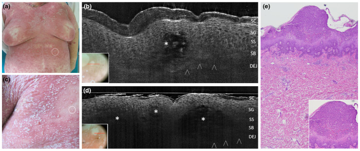
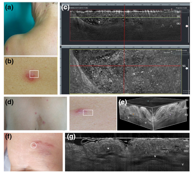
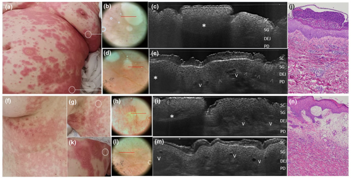
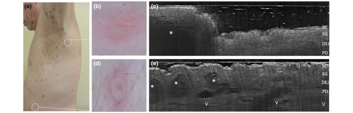
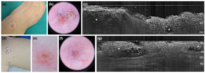
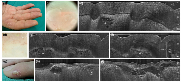
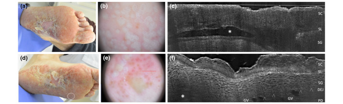
The LC-OCT imaging was able to detect with high accuracy the pustule structure including shape, margins, morphology and cellular content, along with peculiar epidermal and adnexal alterations in each condition, including: Acute Generalized Exanthematous Pustulosis, Generalized pustular psoriasis, Generalized pustular figurate erythema, Subcorneal Pustular Dermatosis, Intraepidermal IgA pustulosis, Palmoplantar pustulosis, Palmoplantar pustular psoriasis. Herpetic whitlow, Acrodermatitis continua of Hallopeau, Vesicopustular Sweet syndrome and Vesicopustular Eosinophilic cellulitis, with pustular appearance, were also compared.
The new LC-OCT can represent a rapid, non-invasive and painless tool which can help differentiating among PSD of different aetiology and microscopic morphology in clinical mimickers in daily practice.
脓疱性皮肤病(PSD)的范围很广,特别具有挑战性,包括炎症性、感染性和非微生物性疾病。此外,尽管脓疱是统一的临床特征,但在疾病的早期阶段,它们可能不存在或未完全发展。线场共聚焦光学相干断层扫描(LC-OCT)是一种最近开发的成像技术,能够对表皮和上部真皮进行非侵入性、体内检查,达到非常高的图像分辨率和虚拟组织学。
我们旨在研究 LC-OCT 在 11 种不同病因、微观特征、身体位置和发病率的 PSD 系列的非侵入性鉴别诊断中的潜力。
对总共 19 名年龄在 35 至 79 岁之间的患者(10 名女性和 9 名男性)进行了完整的 LC-OCT 成像(即 2D/3D 帧、视频)。图像进行了盲法评估,并与相应的组织病理学发现进行了比较。
LC-OCT 成像能够非常准确地检测脓疱结构,包括形状、边缘、形态和细胞内容物,以及每种情况下独特的表皮和附属器改变,包括:急性全身性发疹性脓疱病、全身性脓疱性银屑病、全身性脓疱性类银屑病、亚表皮脓疱性皮肤病、表皮内 IgA 脓疱病、掌跖脓疱病、掌跖脓疱性银屑病。疱疹性白喉、Hallopeau 连续性肢端皮炎、水疱脓疱性Sweet 综合征和水疱脓疱性嗜酸性细胞增多性蜂窝织炎也具有脓疱样表现,也进行了比较。
新的 LC-OCT 可以成为一种快速、非侵入性和无痛的工具,可以帮助在日常实践中区分不同病因和微观形态的 PSD。