Instituto do Coração-Divisão de Pneumologia, Universidade de São Paulo Hospital das Clínicas, Sao Paulo, Brazil
Instituto de Radiologia, Universidade de São Paulo Hospital das Clínicas, Sao Paulo, Brazil.
BMJ Open. 2022 Jun 13;12(6):e059110. doi: 10.1136/bmjopen-2021-059110.
This study aimed to propose a simple, accessible and low-cost predictive clinical model to detect lung lesions due to COVID-19 infection.
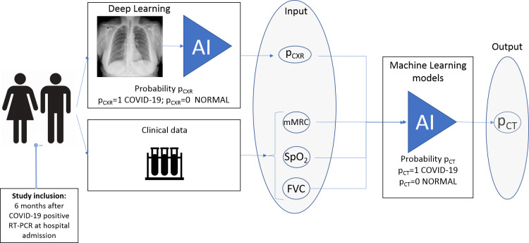
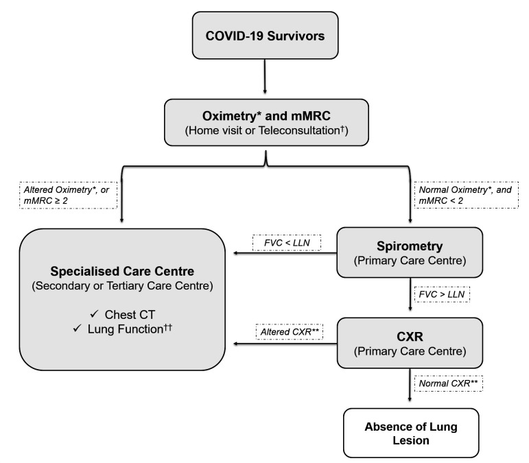
This prospective cohort study included COVID-19 survivors hospitalised between 30 March 2020 and 31 August 2020 followed-up 6 months after hospital discharge. The pulmonary function was assessed using the modified Medical Research Council (mMRC) dyspnoea scale, oximetry (SpO), spirometry (forced vital capacity (FVC)) and chest X-ray (CXR) during an in-person consultation. Patients with abnormalities in at least one of these parameters underwent chest CT. mMRC scale, SpO, FVC and CXR findings were used to build a machine learning model for lung lesion detection on CT.
A tertiary hospital in Sao Paulo, Brazil.
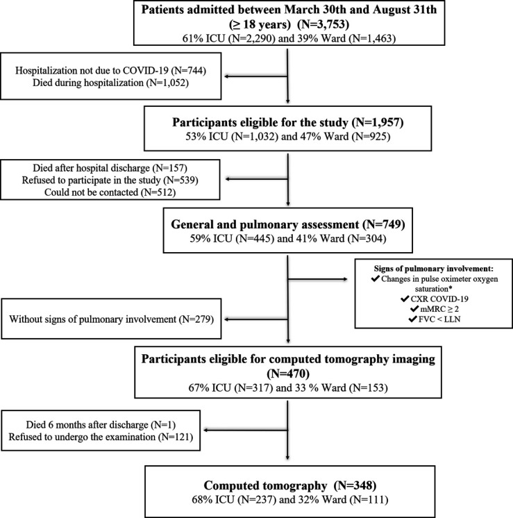
749 eligible RT-PCR-confirmed SARS-CoV-2-infected patients aged ≥18 years.
A predictive clinical model for lung lesion detection on chest CT.
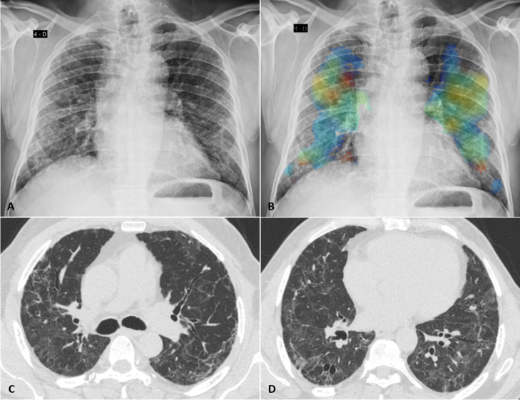
There were 470 patients (63%) that had at least one sign of pulmonary involvement and were eligible for CT. Almost half of them (48%) had significant pulmonary abnormalities, including ground-glass opacities, parenchymal bands, reticulation, traction bronchiectasis and architectural distortion. The machine learning model, including the results of 257 patients with complete data on mMRC, SpO, FVC, CXR and CT, accurately detected pulmonary lesions by the joint data of CXR, mMRC scale, SpO and FVC (sensitivity, 0.85±0.08; specificity, 0.70±0.06; F1-score, 0.79±0.06 and area under the curve, 0.80±0.07).
A predictive clinical model based on CXR, mMRC, oximetry and spirometry data can accurately screen patients with lung lesions after SARS-CoV-2 infection. Given that these examinations are highly accessible and low cost, this protocol can be automated and implemented in different countries for early detection of COVID-19 sequelae.
本研究旨在提出一种简单、易于获取且成本低廉的预测性临床模型,以检测 COVID-19 感染引起的肺部病变。
这项前瞻性队列研究纳入了 2020 年 3 月 30 日至 2020 年 8 月 31 日期间住院的 COVID-19 幸存者,并在出院后 6 个月进行随访。在面对面会诊时,使用改良的医学研究理事会(mMRC)呼吸困难量表、血氧饱和度(SpO)、肺活量计(用力肺活量(FVC))和胸部 X 线(CXR)评估肺功能。至少有一项参数异常的患者进行胸部 CT 检查。使用 mMRC 量表、SpO、FVC 和 CXR 检查结果构建用于 CT 检测肺部病变的机器学习模型。
巴西圣保罗的一家三级医院。
749 名符合条件的 RT-PCR 确诊 SARS-CoV-2 感染患者,年龄≥18 岁。
用于胸部 CT 检测肺部病变的预测性临床模型。
有 470 名(63%)患者至少有一处肺部受累迹象,符合 CT 检查条件。其中近一半(48%)患者存在明显的肺部异常,包括磨玻璃影、实质带、网状影、牵引性支气管扩张和结构扭曲。该机器学习模型纳入了 257 名数据完整的患者(mMRC、SpO、FVC、CXR 和 CT 检查结果均完整),该模型通过 CXR、mMRC 量表、SpO 和 FVC 的联合数据准确地检测到肺部病变(敏感性为 0.85±0.08;特异性为 0.70±0.06;F1 评分为 0.79±0.06;曲线下面积为 0.80±0.07)。
基于 CXR、mMRC、血氧饱和度和肺活量计数据的预测性临床模型可以准确筛查 SARS-CoV-2 感染后肺部病变的患者。鉴于这些检查具有高度可及性且成本低廉,该方案可在不同国家实现自动化并用于 COVID-19 后遗症的早期检测。