Department of Pharmaceutical Sciences and Experimental Therapeutics, The University of Iowa, College of Pharmacy, Iowa City, IA, 52242, USA.
Department of Occupational and Environmental Health, The University of Iowa, College of Public Health, Iowa City, IA, 52242, USA.
Part Fibre Toxicol. 2022 Jun 13;19(1):40. doi: 10.1186/s12989-022-00480-z.
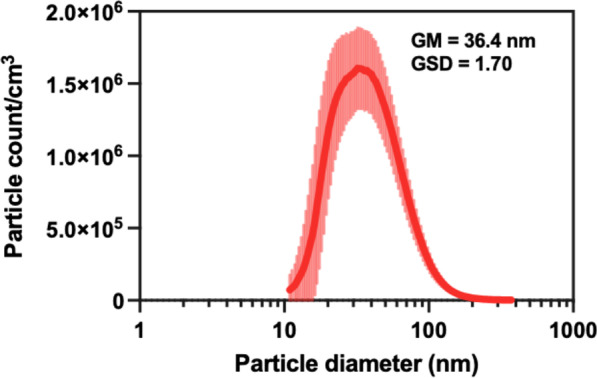
It has been shown that copper oxide nanoparticles (CuO NPs) induce pulmonary toxicity after acute or sub-acute inhalation exposures. However, little is known about the biodistribution and elimination kinetics of inhaled CuO NPs from the respiratory tract. The purposes of this study were to observe the kinetics of pulmonary inflammation during and after CuO NP sub-acute inhalation exposure and to investigate copper (Cu) biodistribution and clearance rate from the exposure site and homeostasis of selected trace elements in secondary organs of BALB/c mice.
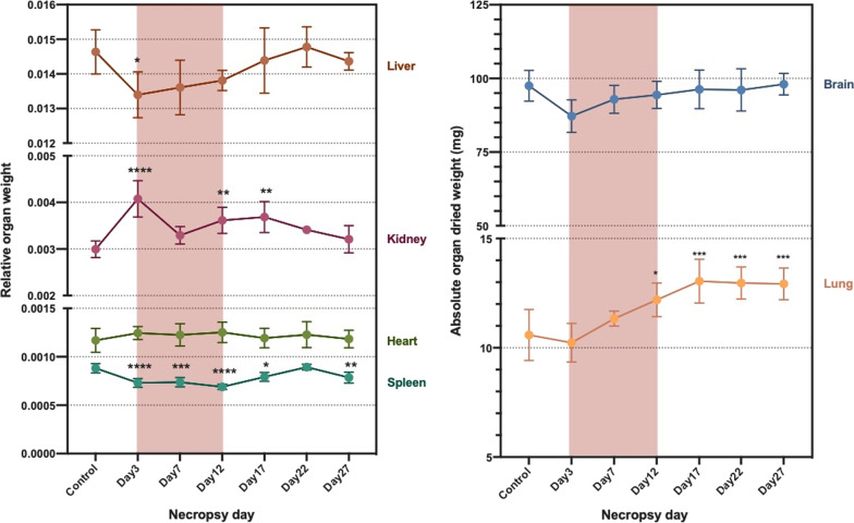
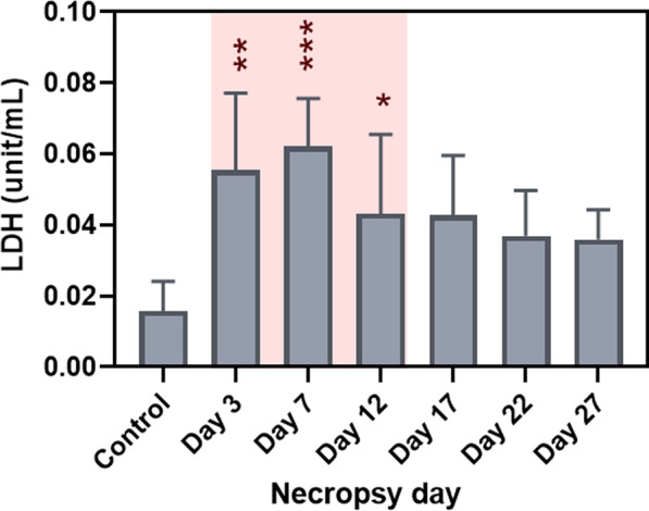
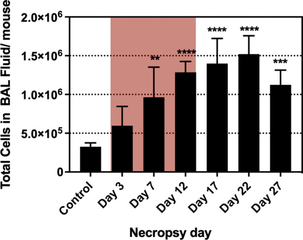
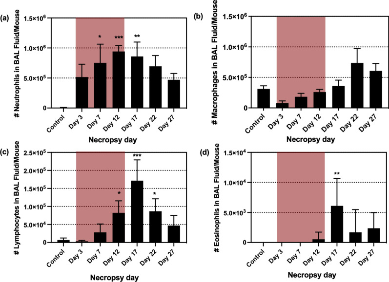
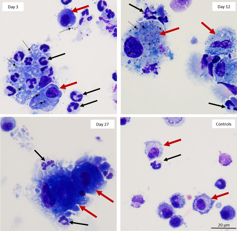
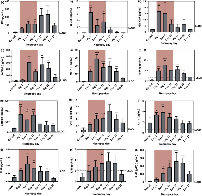
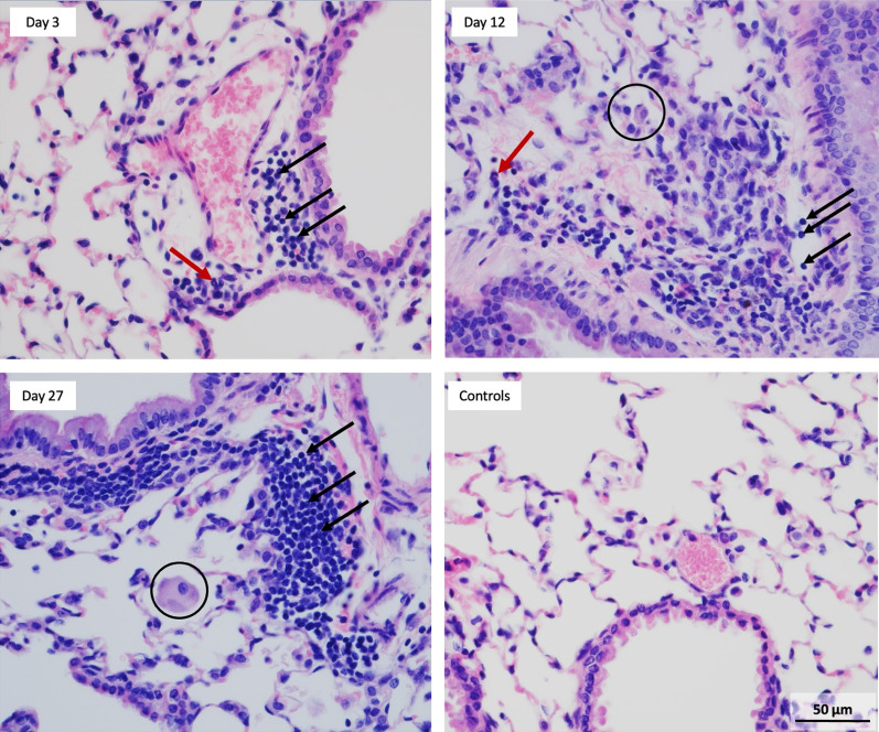
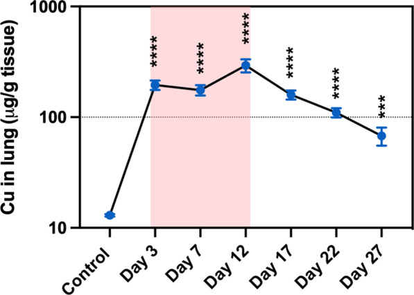
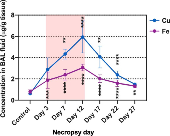
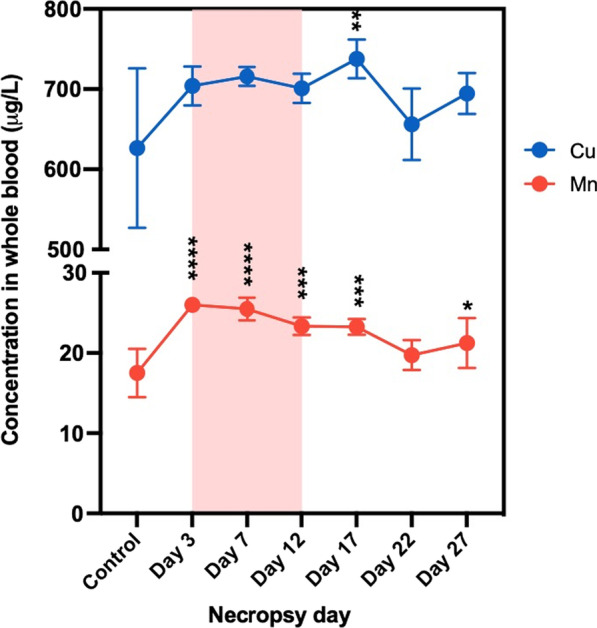
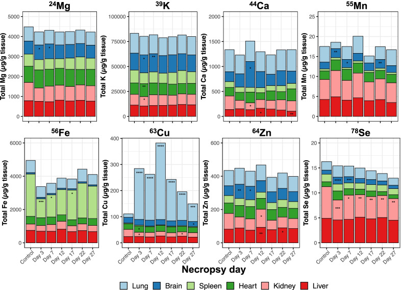
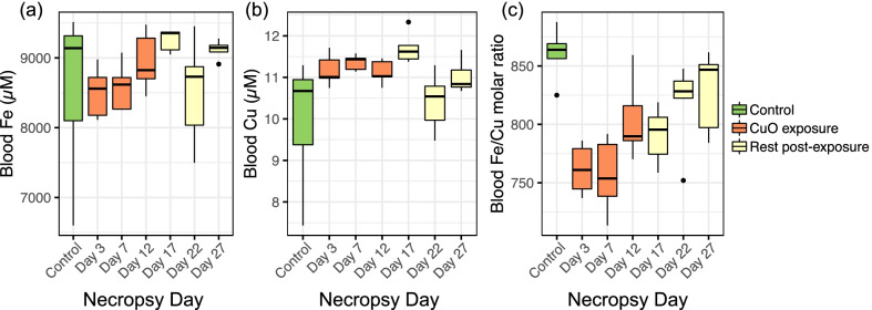
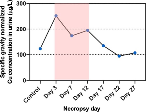
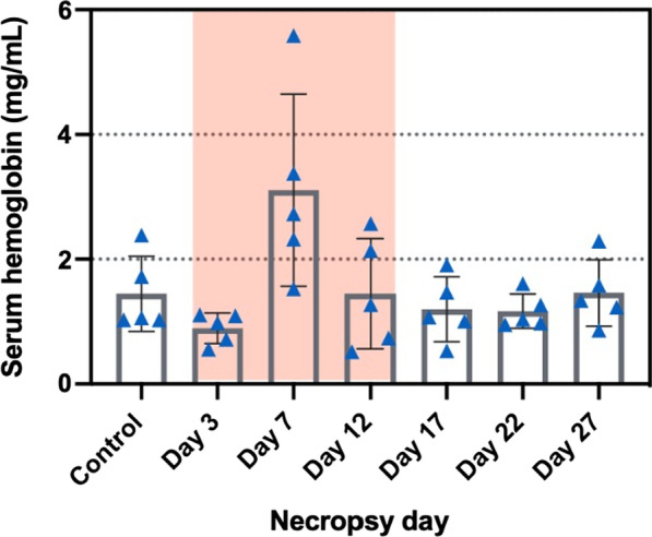
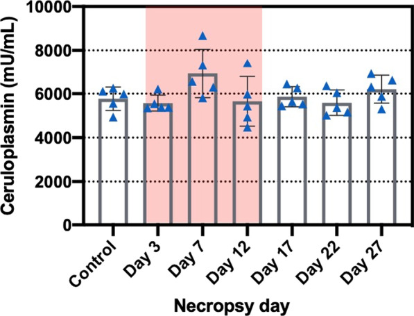
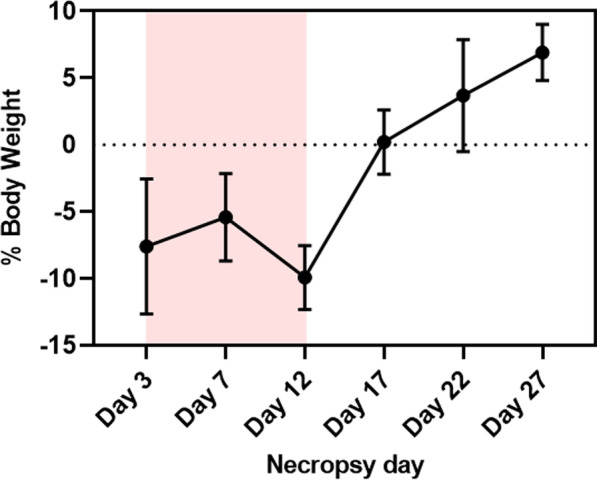
Sub-acute inhalation exposure to CuO NPs led to pulmonary inflammation represented by increases in lactate dehydrogenase, total cell counts, neutrophils, macrophages, inflammatory cytokines, iron levels in bronchoalveolar lavage (BAL) fluid, and lung weight changes. Dosimetry analysis in lung tissues and BAL fluid showed Cu concentration increased steadily during exposure and gradually declined after exposure. Cu elimination from the lung showed first-order kinetics with a half-life of 6.5 days. Total Cu levels were significantly increased in whole blood and heart indicating that inhaled Cu could be translocated into the bloodstream and heart tissue, and potentially have adverse effects on the kidneys and spleen as there were significant changes in the weights of these organs; increase in the kidneys and decrease in the spleen. Furthermore, concentrations of selenium in kidneys and iron in spleen were decreased, pointing to disruption of trace element homeostasis.
Sub-acute inhalation exposure of CuO NPs induced pulmonary inflammation, which was correlated to Cu concentrations in the lungs and started to resolve once exposure ended. Dosimetry analysis showed that Cu in the lungs was translocated into the bloodstream and heart tissue. Secondary organs affected by CuO NPs exposure were kidneys and spleen as they showed the disruption of trace element homeostasis and organ weight changes.
已证明氧化铜纳米颗粒(CuO NPs)在急性或亚急性吸入暴露后会引起肺毒性。然而,对于吸入的 CuO NPs 从呼吸道中的分布和消除动力学知之甚少。本研究的目的是观察 CuO NP 亚急性吸入暴露期间和之后肺部炎症的动力学,并研究铜(Cu)从暴露部位的生物分布和清除率以及 BALB/c 小鼠次级器官中选定痕量元素的内稳态。
CuO NPs 的亚急性吸入暴露导致肺炎症,表现为乳酸脱氢酶、总细胞计数、中性粒细胞、巨噬细胞、炎症细胞因子、支气管肺泡灌洗液(BAL)中铁水平以及肺重量变化增加。肺组织和 BAL 液中的剂量测定分析表明,暴露期间 Cu 浓度稳步增加,暴露后逐渐下降。Cu 从肺部的消除呈一级动力学,半衰期为 6.5 天。全血和心脏中的总 Cu 水平显着升高,表明吸入的 Cu 可以转移到血液和心脏组织中,并可能对肾脏和脾脏产生不利影响,因为这些器官的重量发生了显着变化;肾脏重量增加,脾脏重量减少。此外,肾脏中的硒浓度和脾脏中的铁浓度降低,表明微量元素内稳态受到干扰。
CuO NPs 的亚急性吸入暴露会引起肺部炎症,这与肺部的 Cu 浓度相关,一旦暴露结束,炎症就会开始消退。剂量测定分析表明,肺部的 Cu 被转移到血液和心脏组织中。受 CuO NPs 暴露影响的次级器官是肾脏和脾脏,因为它们表现出微量元素内稳态和器官重量变化的破坏。