Department of Neurosciences, Lerner Research Institute, Cleveland Clinic Foundation, and Case Western Reserve University, 9500 Euclid Avenue, Cleveland, OH, 44195, USA.
Hooke Laboratories, Inc., Lawrence, MA, USA.
Acta Neuropathol Commun. 2022 Jun 15;10(1):87. doi: 10.1186/s40478-022-01391-y.
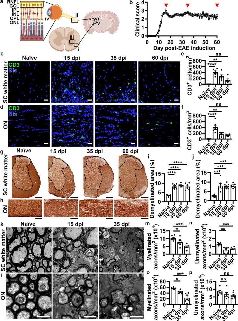
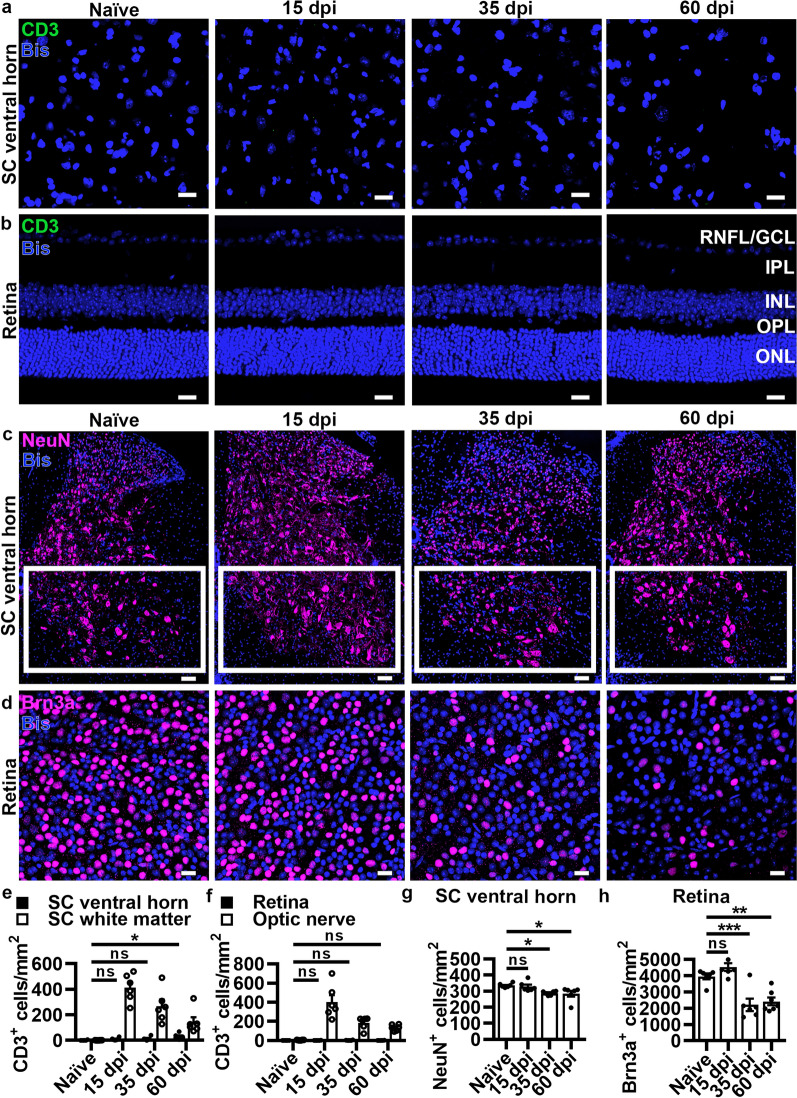
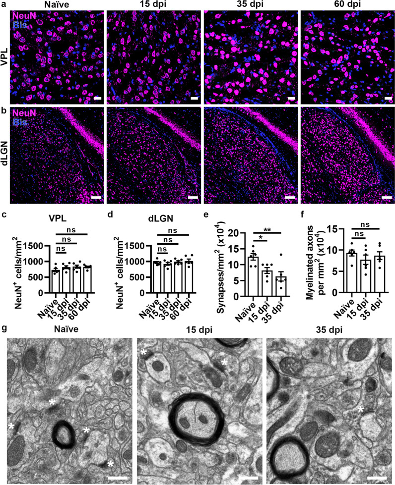
Thalamic volume is associated with clinical disability in multiple sclerosis (MS) and is vulnerable to secondary neurodegeneration due to its extensive connectivity throughout the central nervous system (CNS). Using a model of autoimmune demyelination that exhibits CNS-infiltrating immune cells in both spinal cord white matter and optic nerve, we sought to evaluate neurodegenerative changes due to lesions affecting the spino- and retino-thalamic pathways. We found comparable axonal loss in spinal cord white matter and optic nerve during the acute phase of disease consistent with synaptic loss, but not neuronal cell body loss in the thalamic nuclei that receive input from these discrete pathways. Loss of spinal cord neurons or retinal ganglion cells retrograde to their respective axons was not observed until the chronic phase of disease, where optical coherence tomography (OCT) documented reduced inner retinal thickness. In patients with relapsing-remitting MS without a history of optic neuritis, OCT measures of inner retinal volume correlated with retino-thalamic (lateral geniculate nucleus) and spino-thalamic (ventral posterior nucleus) volume as well as neuroperformance measures. These data suggest retinal imaging may serve as an important noninvasive predictor of neurodegeneration in MS.
丘脑体积与多发性硬化症(MS)的临床残疾有关,由于其与中枢神经系统(CNS)的广泛连接,易受到继发神经退行性变的影响。我们使用一种自身免疫性脱髓鞘模型,该模型在脊髓白质和视神经中均表现出中枢神经系统浸润的免疫细胞,旨在评估因影响脊髓和视丘束通路的病变引起的神经退行性变化。我们发现在疾病的急性期,脊髓白质和视神经中的轴突丢失相当,这与突触丢失一致,但接收这些离散通路输入的丘脑核中的神经元细胞体丢失并不明显。直到疾病的慢性期,才观察到脊髓神经元或视网膜神经节细胞向其各自轴突的逆行性丧失,此时光学相干断层扫描(OCT)记录到内视网膜厚度降低。在没有视神经炎病史的复发缓解型 MS 患者中,OCT 对内视网膜体积的测量与视丘束(外侧膝状体核)和脊髓束(腹后核)体积以及神经功能测量相关。这些数据表明,视网膜成像可能成为 MS 中神经退行性变的重要无创预测指标。