Division of Medical Genetics, Department of Internal Medicine, McGovern Medical School The University of Texas Health Science Center at Houston (A.C., P.G., S.M., K.K., Z.Z., S.K.P.' A.K., C.S.K., D.M.M.).
Graduate School of Biomedical Sciences, University of Texas MD Anderson Cancer Center and UTHealth, Houston (P.G.).
Arterioscler Thromb Vasc Biol. 2022 Aug;42(8):1005-1022. doi: 10.1161/ATVBAHA.121.317451. Epub 2022 Jun 16.
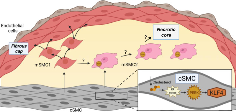
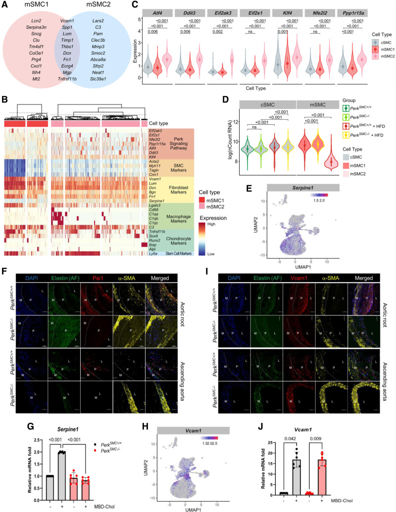
Vascular smooth muscle cells (SMCs) undergo complex phenotypic modulation with atherosclerotic plaque formation in hyperlipidemic mice, which is characterized by de-differentiation and heterogeneous increases in the expression of macrophage, fibroblast, osteogenic, and stem cell markers. An increase of cellular cholesterol in SMCs triggers similar phenotypic changes in vitro with exposure to free cholesterol due to cholesterol entering the endoplasmic reticulum, triggering endoplasmic reticulum stress and activating Perk (protein kinase RNA-like endoplasmic reticulum kinase) signaling.
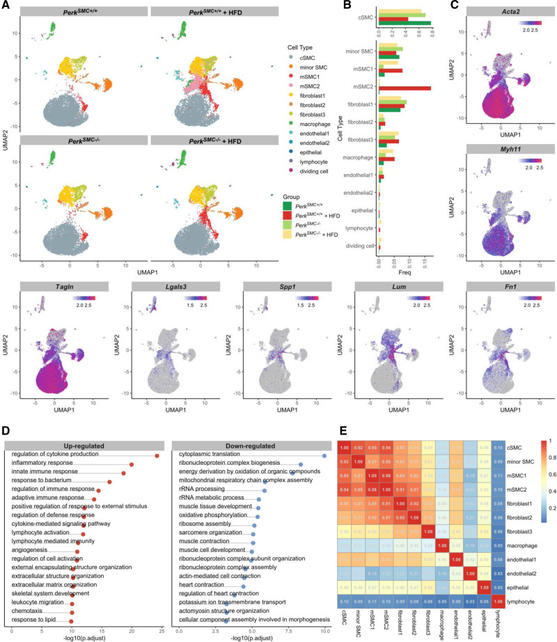
We generated an SMC-specific knockout mouse model, induced hyperlipidemia in the mice by AAV- injection, and subjected them to a high-fat diet. We then assessed atherosclerotic plaque formation and performed single-cell transcriptomic studies using aortic tissue from these mice.
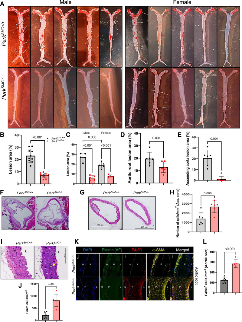
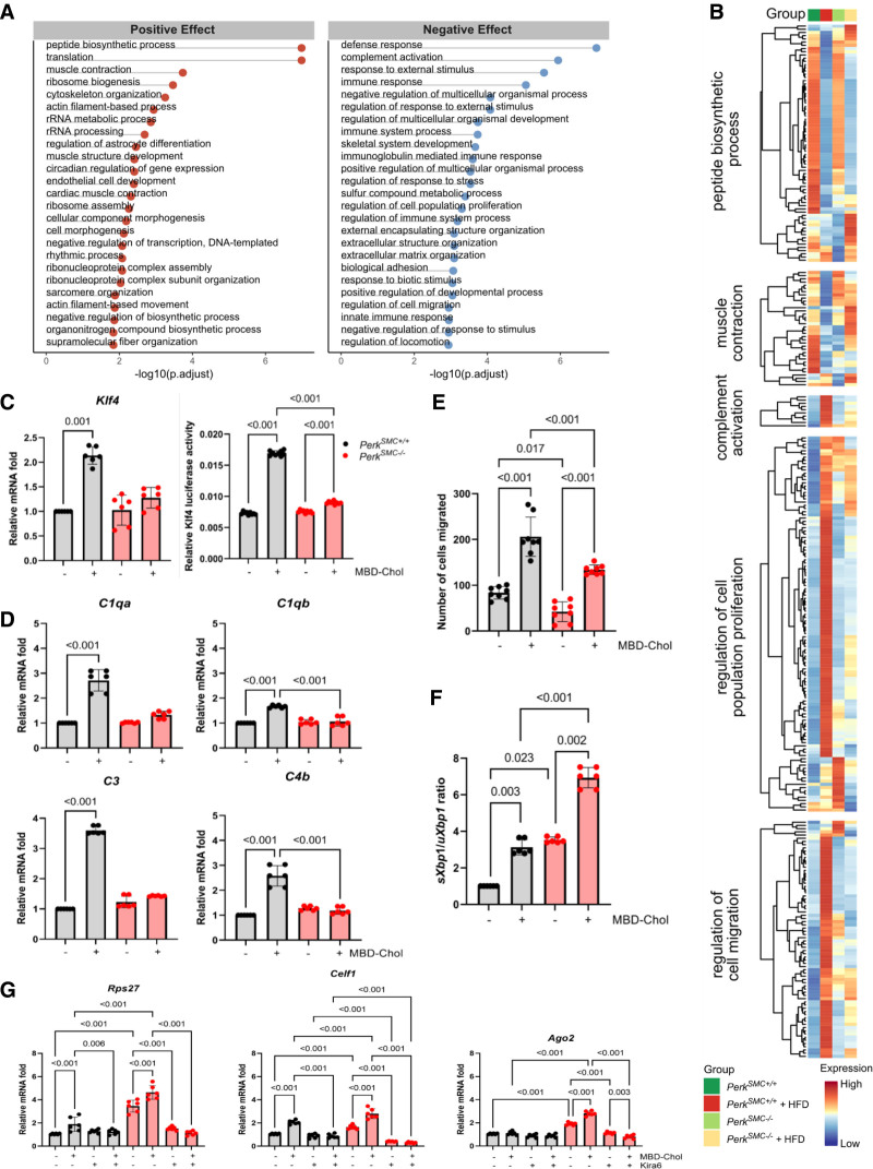
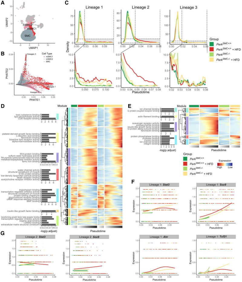
SMC-specific deletion of Perk reduces atherosclerotic plaque formation in male hyperlipidemic mice by 80%. Single-cell transcriptomic data identify 2 clusters of modulated SMCs in hyperlipidemic mice, one of which is absent when is deleted in SMCs. The 2 modulated SMC clusters have significant overlap of transcriptional changes, but the Perk-dependent cluster uniquely shows a global decrease in the number of transcripts. SMC-specific Perk deletion also prevents migration of both contractile and modulated SMCs from the medial layer of the aorta.
Our results indicate that hypercholesterolemia drives both Perk-dependent and Perk-independent SMC modulation and that deficiency of Perk significantly blocks atherosclerotic plaque formation.
在高脂血症小鼠的动脉粥样硬化斑块形成过程中,血管平滑肌细胞(SMCs)经历复杂的表型调节,表现为去分化和巨噬细胞、成纤维细胞、成骨细胞和干细胞标志物表达的异质性增加。SMC 中细胞胆固醇的增加会导致类似的表型变化,因为胆固醇进入内质网,触发内质网应激并激活 Perk(蛋白激酶 RNA 样内质网激酶)信号通路,使 SMC 暴露于游离胆固醇。
我们构建了 SMC 特异性 Perk 敲除小鼠模型,通过 AAV-注射诱导小鼠发生高脂血症,并让它们接受高脂肪饮食。然后,我们评估了这些小鼠主动脉组织中的动脉粥样硬化斑块形成情况,并进行了单细胞转录组学研究。
SMC 特异性 Perk 缺失可使雄性高脂血症小鼠的动脉粥样硬化斑块形成减少 80%。单细胞转录组学数据鉴定出高脂血症小鼠中存在 2 种调节型 SMC 簇,当 SMC 中缺失时,其中一种簇就不存在了。这 2 种调节型 SMC 簇具有显著重叠的转录变化,但依赖 Perk 的簇中独特地表现出转录本数量的全面减少。SMC 特异性 Perk 缺失还可防止收缩型和调节型 SMC 从中膜层迁移到主动脉。
我们的研究结果表明,高胆固醇血症可驱动 Perk 依赖性和非依赖性 SMC 调节,而 Perk 缺乏可显著阻止动脉粥样硬化斑块的形成。