• 文献检索
  • 文档翻译
  • 深度研究
  • 学术资讯
  • Suppr Zotero 插件Zotero 插件
  • 邀请有礼
  • 套餐&价格
  • 历史记录
应用&插件
Suppr Zotero 插件Zotero 插件浏览器插件Mac 客户端Windows 客户端微信小程序
定价
高级版会员购买积分包购买API积分包
服务
文献检索文档翻译深度研究API 文档MCP 服务
关于我们
关于 Suppr公司介绍联系我们用户协议隐私条款
关注我们

Suppr 超能文献

核心技术专利:CN118964589B侵权必究
粤ICP备2023148730 号-1Suppr @ 2026

文献检索

告别复杂PubMed语法,用中文像聊天一样搜索,搜遍4000万医学文献。AI智能推荐,让科研检索更轻松。

立即免费搜索

文件翻译

保留排版,准确专业,支持PDF/Word/PPT等文件格式,支持 12+语言互译。

免费翻译文档

深度研究

AI帮你快速写综述,25分钟生成高质量综述,智能提取关键信息,辅助科研写作。

立即免费体验

通过全原子分子动力学模拟突触融合蛋白-SNARE 复合蛋白桥接小泡和平面脂双层。

All-atom molecular dynamics simulations of Synaptotagmin-SNARE-complexin complexes bridging a vesicle and a flat lipid bilayer.

机构信息

Department of Biophysics, The University of Texas Southwestern Medical Center, Dallas, United States.

Department of Biochemistry, University of Texas Southwestern Medical Center, Dallas, United States.

出版信息

Elife. 2022 Jun 16;11:e76356. doi: 10.7554/eLife.76356.

DOI:10.7554/eLife.76356
PMID:35708237
原文链接:https://pmc.ncbi.nlm.nih.gov/articles/PMC9239685/
Abstract

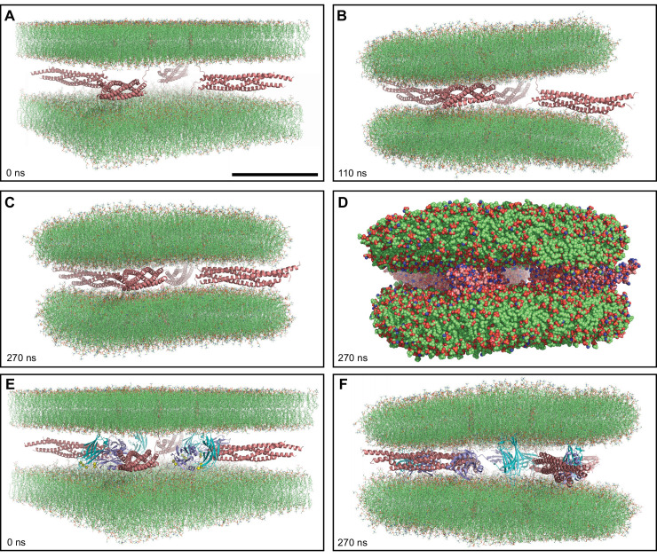
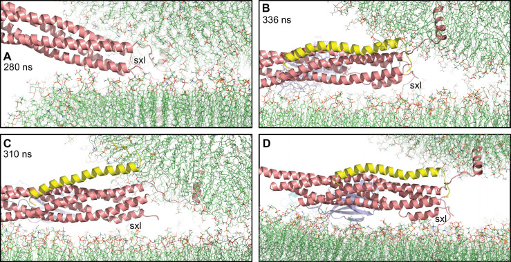
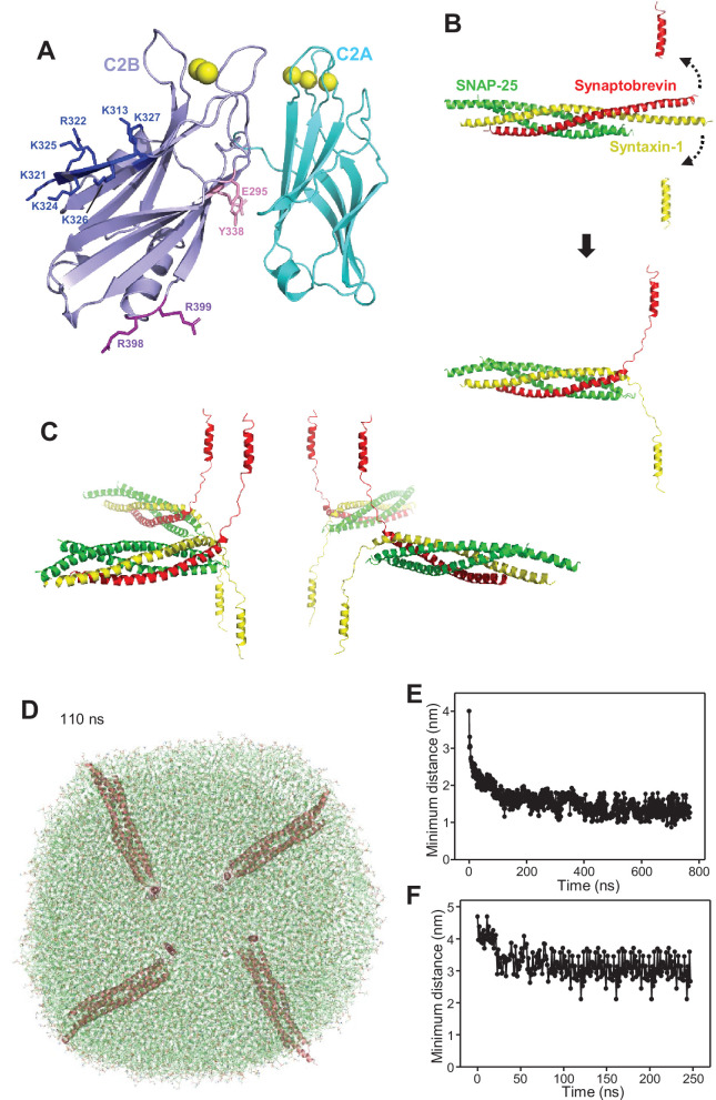
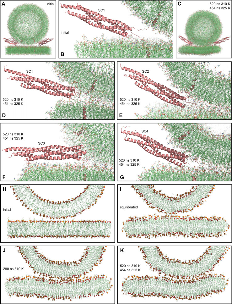
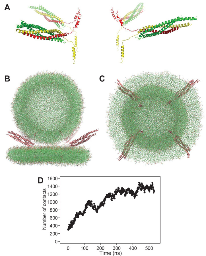
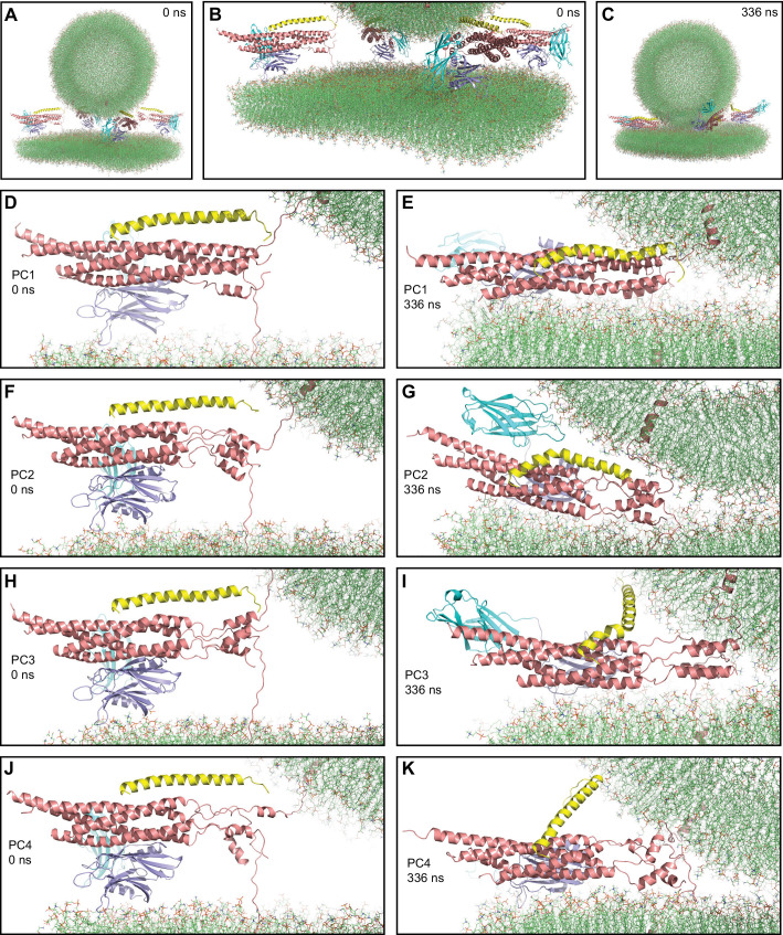
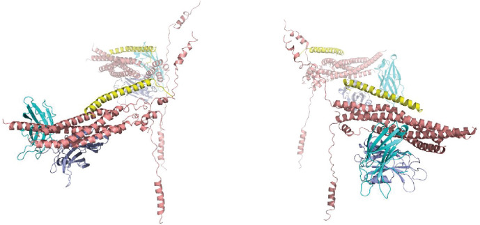
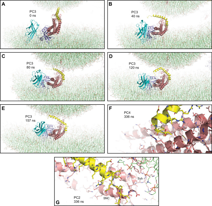
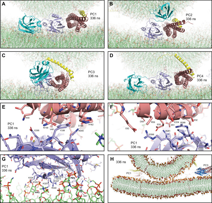
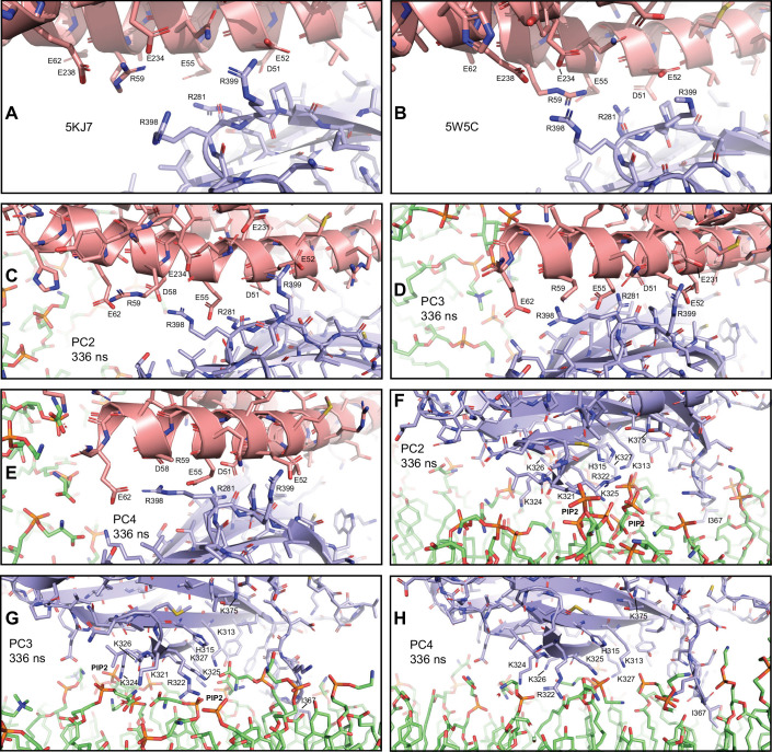
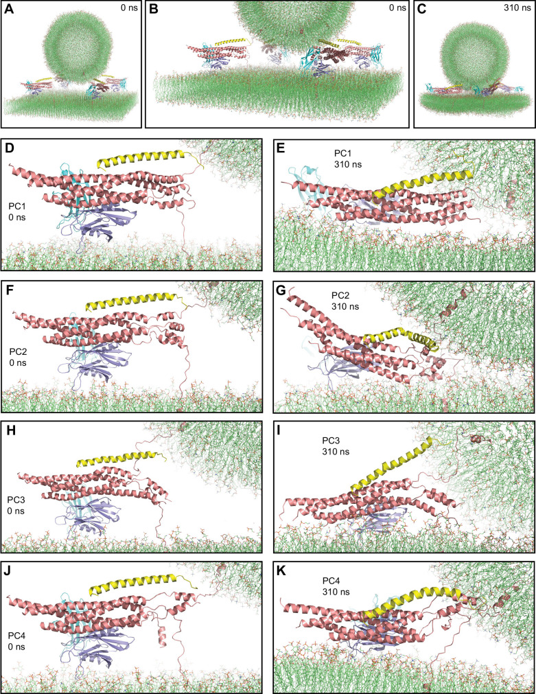
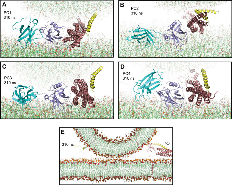
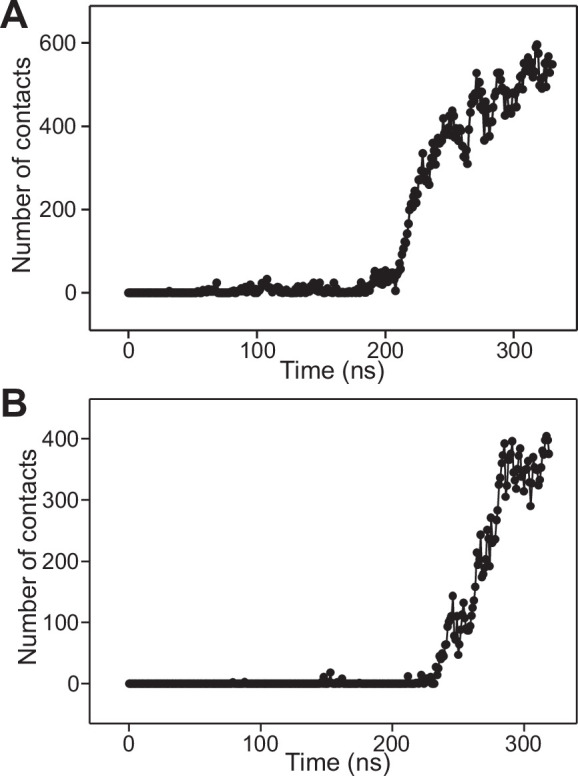
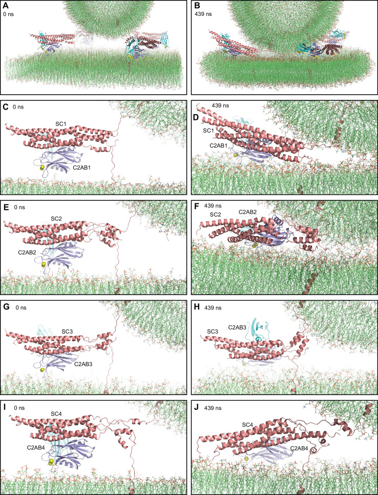
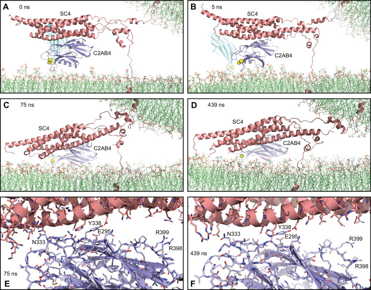
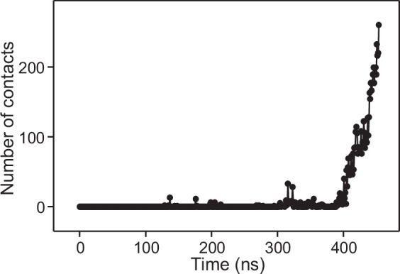
Synaptic vesicles are primed into a state that is ready for fast neurotransmitter release upon Ca-binding to Synaptotagmin-1. This state likely includes trans-SNARE complexes between the vesicle and plasma membranes that are bound to Synaptotagmin-1 and complexins. However, the nature of this state and the steps leading to membrane fusion are unclear, in part because of the difficulty of studying this dynamic process experimentally. To shed light into these questions, we performed all-atom molecular dynamics simulations of systems containing trans-SNARE complexes between two flat bilayers or a vesicle and a flat bilayer with or without fragments of Synaptotagmin-1 and/or complexin-1. Our results need to be interpreted with caution because of the limited simulation times and the absence of key components, but suggest mechanistic features that may control release and help visualize potential states of the primed Synaptotagmin-1-SNARE-complexin-1 complex. The simulations suggest that SNAREs alone induce formation of extended membrane-membrane contact interfaces that may fuse slowly, and that the primed state contains macromolecular assemblies of trans-SNARE complexes bound to the Synaptotagmin-1 CB domain and complexin-1 in a spring-loaded configuration that prevents premature membrane merger and formation of extended interfaces, but keeps the system ready for fast fusion upon Ca influx.

摘要

当钙离子与突触融合蛋白 1(Synaptotagmin-1)结合时,突触囊泡被预先置于一种准备快速释放神经递质的状态。这种状态可能包括与突触融合蛋白 1 和衔接蛋白结合的囊泡和质膜之间的跨 SNARE 复合物。然而,这种状态的本质以及导致膜融合的步骤尚不清楚,部分原因是由于实验研究这一动态过程的难度。为了阐明这些问题,我们对包含两个平面双层或一个囊泡和一个平面双层之间的跨 SNARE 复合物的系统进行了全原子分子动力学模拟,其中包含或不包含突触融合蛋白 1 和/或衔接蛋白 1 的片段。由于模拟时间有限且缺乏关键成分,我们的结果需要谨慎解释,但它们提出了可能控制释放的机械特征,并有助于可视化预先形成的突触融合蛋白 1-SNARE-衔接蛋白 1 复合物的潜在状态。模拟表明,SNARE 蛋白本身会诱导形成扩展的膜-膜接触界面,这些界面可能会缓慢融合,而预先形成的状态包含与突触融合蛋白 1 CB 结构域结合的跨 SNARE 复合物的大分子组装体和衔接蛋白 1,处于弹簧加载配置中,这种配置可防止过早的膜融合和扩展界面的形成,但使系统在钙离子流入时能够快速融合。

https://cdn.ncbi.nlm.nih.gov/pmc/blobs/5571/9239685/467b00377925/elife-76356-fig6.jpg
https://cdn.ncbi.nlm.nih.gov/pmc/blobs/5571/9239685/416da9dd7f7a/elife-76356-fig1.jpg
https://cdn.ncbi.nlm.nih.gov/pmc/blobs/5571/9239685/9ff57084ae06/elife-76356-fig1-figsupp1.jpg
https://cdn.ncbi.nlm.nih.gov/pmc/blobs/5571/9239685/7e5c3598083d/elife-76356-fig2.jpg
https://cdn.ncbi.nlm.nih.gov/pmc/blobs/5571/9239685/46e431825a87/elife-76356-fig2-figsupp1.jpg
https://cdn.ncbi.nlm.nih.gov/pmc/blobs/5571/9239685/dd57d1f27f5a/elife-76356-fig3.jpg
https://cdn.ncbi.nlm.nih.gov/pmc/blobs/5571/9239685/fa1729d348c9/elife-76356-fig3-figsupp1.jpg
https://cdn.ncbi.nlm.nih.gov/pmc/blobs/5571/9239685/3dfcfc4c2600/elife-76356-fig3-figsupp2.jpg
https://cdn.ncbi.nlm.nih.gov/pmc/blobs/5571/9239685/233032303fca/elife-76356-fig4.jpg
https://cdn.ncbi.nlm.nih.gov/pmc/blobs/5571/9239685/896ac31f601f/elife-76356-fig4-figsupp1.jpg
https://cdn.ncbi.nlm.nih.gov/pmc/blobs/5571/9239685/ecf0bd0888e3/elife-76356-fig4-figsupp2.jpg
https://cdn.ncbi.nlm.nih.gov/pmc/blobs/5571/9239685/8e8aedca6d83/elife-76356-fig4-figsupp3.jpg
https://cdn.ncbi.nlm.nih.gov/pmc/blobs/5571/9239685/414ea2a2fd1c/elife-76356-fig4-figsupp4.jpg
https://cdn.ncbi.nlm.nih.gov/pmc/blobs/5571/9239685/ffb29e51fe9b/elife-76356-fig5.jpg
https://cdn.ncbi.nlm.nih.gov/pmc/blobs/5571/9239685/e0ec62913c30/elife-76356-fig5-figsupp1.jpg
https://cdn.ncbi.nlm.nih.gov/pmc/blobs/5571/9239685/76c89f66e1cb/elife-76356-fig5-figsupp2.jpg
https://cdn.ncbi.nlm.nih.gov/pmc/blobs/5571/9239685/467b00377925/elife-76356-fig6.jpg

相似文献

1
All-atom molecular dynamics simulations of Synaptotagmin-SNARE-complexin complexes bridging a vesicle and a flat lipid bilayer.通过全原子分子动力学模拟突触融合蛋白-SNARE 复合蛋白桥接小泡和平面脂双层。
Elife. 2022 Jun 16;11:e76356. doi: 10.7554/eLife.76356.
2
A complexin/synaptotagmin 1 switch controls fast synaptic vesicle exocytosis.一种复合体蛋白/突触结合蛋白1开关控制快速突触小泡胞吐作用。
Cell. 2006 Sep 22;126(6):1175-87. doi: 10.1016/j.cell.2006.08.030.
3
The primed SNARE-complexin-synaptotagmin complex for neuronal exocytosis.用于神经元胞吐作用的引发型SNARE-结合蛋白-突触结合蛋白复合物
Nature. 2017 Aug 24;548(7668):420-425. doi: 10.1038/nature23484. Epub 2017 Aug 16.
4
Synergistic roles of Synaptotagmin-1 and complexin in calcium-regulated neuronal exocytosis.突触结合蛋白 1 和复合蛋白在钙调节的神经元胞吐中的协同作用。
Elife. 2020 May 13;9:e54506. doi: 10.7554/eLife.54506.
5
Synaptotagmin 1 clamps synaptic vesicle fusion in mammalian neurons independent of complexin.突触结合蛋白 1 通过独立于钙联蛋白的方式在哺乳神经元中钳制突触囊泡融合。
Nat Commun. 2019 Sep 9;10(1):4076. doi: 10.1038/s41467-019-12015-w.
6
Subtle Interplay between synaptotagmin and complexin binding to the SNARE complex.突触融合蛋白和复合蛋白与 SNARE 复合物之间的微妙相互作用。
J Mol Biol. 2013 Sep 23;425(18):3461-75. doi: 10.1016/j.jmb.2013.07.001. Epub 2013 Jul 9.
7
Concurrent binding of complexin and synaptotagmin to liposome-embedded SNARE complexes.复合体蛋白和突触结合蛋白与脂质体包埋的SNARE复合体的同时结合。
Biochemistry. 2009 Feb 3;48(4):657-9. doi: 10.1021/bi801962d.
8
Munc18a does not alter fusion rates mediated by neuronal SNAREs, synaptotagmin, and complexin.Munc18a不会改变由神经元SNARE蛋白、突触结合蛋白和复合体蛋白介导的融合速率。
J Biol Chem. 2015 Apr 17;290(16):10518-34. doi: 10.1074/jbc.M114.630772. Epub 2015 Feb 25.
9
synaptotagmin mutants reveal essential functions for the C2B domain in Ca2+-triggered fusion and recycling of synaptic vesicles in vivo.突触结合蛋白突变体揭示了C2B结构域在体内Ca2+触发的突触小泡融合和循环中的重要功能。
J Neurosci. 2001 Mar 1;21(5):1421-33. doi: 10.1523/JNEUROSCI.21-05-01421.2001.
10
Binding of the complexin N terminus to the SNARE complex potentiates synaptic-vesicle fusogenicity.复合蛋白 N 端与 SNARE 复合物的结合增强了突触囊泡的融合能力。
Nat Struct Mol Biol. 2010 May;17(5):568-75. doi: 10.1038/nsmb.1791. Epub 2010 Apr 18.

引用本文的文献

1
Structural insights into lipid membrane binding by human ferlins.人类ferlins蛋白与脂质膜结合的结构见解
EMBO J. 2025 May 28. doi: 10.1038/s44318-025-00463-8.
2
Evaluation of synaptotagmin-1 action models by all-atom molecular dynamics simulations.通过全原子分子动力学模拟评估突触结合蛋白-1作用模型。
FEBS Open Bio. 2025 May;15(5):699-713. doi: 10.1002/2211-5463.13966. Epub 2025 Jan 15.
3
Molecular Dynamics Simulation for Membrane Fusion.膜融合的分子动力学模拟

本文引用的文献

1
Molecular Mechanisms Underlying Neurotransmitter Release.神经递质释放的分子机制。
Annu Rev Biophys. 2022 May 9;51:377-408. doi: 10.1146/annurev-biophys-111821-104732. Epub 2022 Feb 15.
2
Munc13 structural transitions and oligomers that may choreograph successive stages in vesicle priming for neurotransmitter release.Munc13 结构的转变和寡聚体可能在神经递质释放的囊泡引发的连续阶段中进行协调。
Proc Natl Acad Sci U S A. 2022 Feb 15;119(7). doi: 10.1073/pnas.2121259119.
3
Control of neurotransmitter release by two distinct membrane-binding faces of the Munc13-1 CCB region.
Methods Mol Biol. 2025;2887:53-68. doi: 10.1007/978-1-0716-4314-3_3.
4
A lever hypothesis for Synaptotagmin-1 action in neurotransmitter release.关于突触结合蛋白-1在神经递质释放中作用的杠杆假说。
Proc Natl Acad Sci U S A. 2025 Jan 7;122(1):e2417941121. doi: 10.1073/pnas.2417941121. Epub 2024 Dec 30.
5
Neurotransmitter release is triggered by a calcium-induced rearrangement in the Synaptotagmin-1/SNARE complex primary interface.神经递质释放是由突触融合蛋白 1/SNARE 复合物主要界面中钙诱导的重排触发的。
Proc Natl Acad Sci U S A. 2024 Oct 15;121(42):e2409636121. doi: 10.1073/pnas.2409636121. Epub 2024 Oct 7.
6
Dynamic formation of the protein-lipid prefusion complex.蛋白质-脂类预融合复合物的动态形成。
Biophys J. 2024 Oct 15;123(20):3569-3586. doi: 10.1016/j.bpj.2024.09.009. Epub 2024 Sep 10.
7
Exploring the structural dynamics of the vesicle priming machinery.探索囊泡引发机制的结构动力学。
Biochem Soc Trans. 2024 Aug 28;52(4):1715-1725. doi: 10.1042/BST20231333.
8
Neurotransmitter release is triggered by a calcium-induced rearrangement in the Synaptotagmin-1/SNARE complex primary interface.神经递质的释放是由钙诱导的突触结合蛋白-1/可溶性N-乙基马来酰胺敏感因子附着蛋白受体(SNARE)复合体初级界面重排所触发的。
bioRxiv. 2024 Jun 21:2024.06.17.599435. doi: 10.1101/2024.06.17.599435.
9
A lever hypothesis for Synaptotagmin-1 action in neurotransmitter release.关于突触结合蛋白-1在神经递质释放中作用的杠杆假说。
bioRxiv. 2024 Jun 18:2024.06.17.599417. doi: 10.1101/2024.06.17.599417.
10
Coarse-grained modeling of annexin A2-induced microdomain formation on a vesicle.网格蛋白包被小泡形成过程中 annexin A2 诱导的微区的粗粒化建模。
Biophys J. 2024 Aug 20;123(16):2431-2442. doi: 10.1016/j.bpj.2024.06.006. Epub 2024 Jun 10.
Munc13-1 CCB 区域的两个不同膜结合面控制神经递质释放。
Elife. 2021 Nov 15;10:e72030. doi: 10.7554/eLife.72030.
4
Evaluation of the tert-butyl group as a probe for NMR studies of macromolecular complexes.评价叔丁基作为核磁共振研究大分子配合物的探针。
J Biomol NMR. 2021 Sep;75(8-9):347-363. doi: 10.1007/s10858-021-00380-y. Epub 2021 Sep 9.
5
Tight docking of membranes before fusion represents a metastable state with unique properties.在融合之前,膜的紧密对接代表了一种具有独特性质的亚稳态。
Nat Commun. 2021 Jun 14;12(1):3606. doi: 10.1038/s41467-021-23722-8.
6
Nascent fusion pore opening monitored at single-SNAREpin resolution.初生融合孔在单 SNARE 蛋白分辨率下的开启监测。
Proc Natl Acad Sci U S A. 2021 Feb 2;118(5). doi: 10.1073/pnas.2024922118.
7
Symmetrical arrangement of proteins under release-ready vesicles in presynaptic terminals.突触前末梢中准备释放的囊泡下蛋白质的对称排列。
Proc Natl Acad Sci U S A. 2021 Feb 2;118(5). doi: 10.1073/pnas.2024029118.
8
SNARE complex alters the interactions of the Ca sensor synaptotagmin 1 with lipid bilayers.SNARE 复合物改变了钙传感器突触融合蛋白 1 与脂双层的相互作用。
Biophys J. 2021 Feb 16;120(4):642-661. doi: 10.1016/j.bpj.2020.12.025. Epub 2021 Jan 14.
9
Ca-dependent release of synaptotagmin-1 from the SNARE complex on phosphatidylinositol 4,5-bisphosphate-containing membranes.钙离子依赖的突触融合蛋白-1从含有磷酸肌醇 4,5-二磷酸的 SNARE 复合物中的释放。
Elife. 2020 Aug 18;9:e57154. doi: 10.7554/eLife.57154.
10
Complexin Suppresses Spontaneous Exocytosis by Capturing the Membrane-Proximal Regions of VAMP2 and SNAP25.复合蛋白通过捕获 VAMP2 和 SNAP25 的膜近端区域来抑制自发性胞吐作用。
Cell Rep. 2020 Jul 21;32(3):107926. doi: 10.1016/j.celrep.2020.107926.