Department of Cell Biology, Yale University School of Medicine, New Haven, CT 06520.
Department of Microbial Pathogenesis, Yale University School of Medicine, New Haven, CT 06520.
Proc Natl Acad Sci U S A. 2021 Feb 2;118(5). doi: 10.1073/pnas.2024029118.
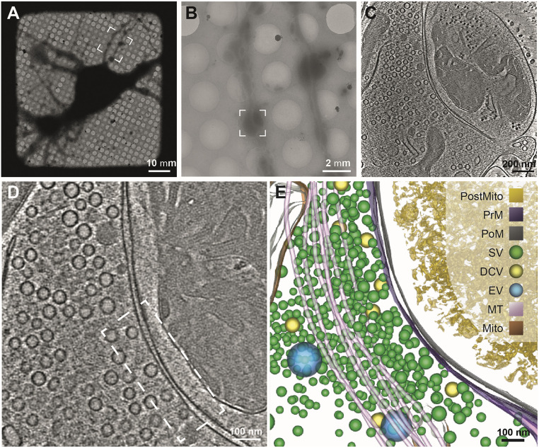
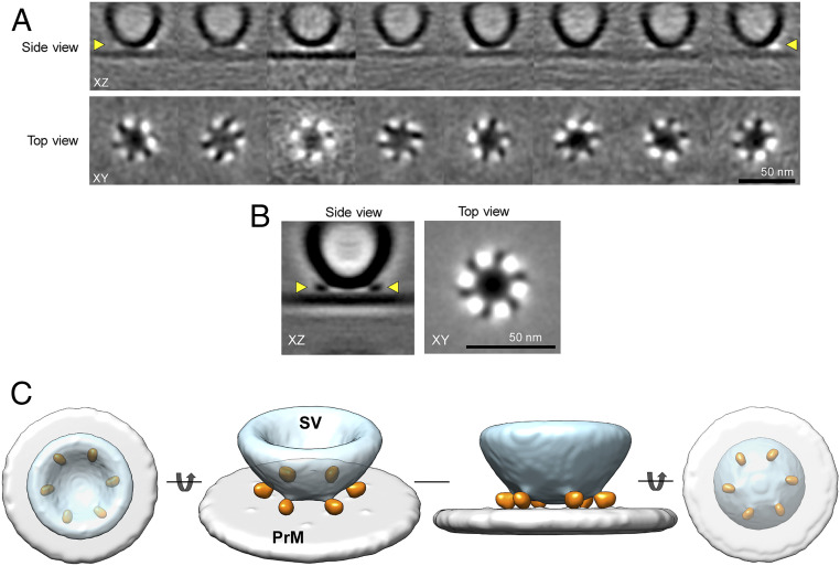
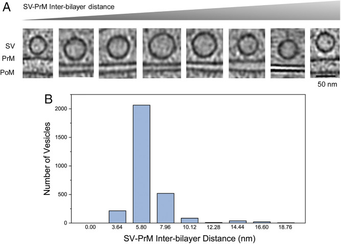
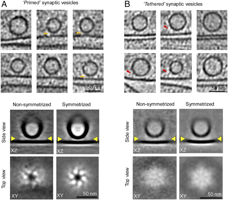
Controlled release of neurotransmitters stored in synaptic vesicles (SVs) is a fundamental process that is central to all information processing in the brain. This relies on tight coupling of the SV fusion to action potential-evoked presynaptic Ca influx. This Ca-evoked release occurs from a readily releasable pool (RRP) of SVs docked to the plasma membrane (PM). The protein components involved in initial SV docking/tethering and the subsequent priming reactions which make the SV release ready are known. Yet, the supramolecular architecture and sequence of molecular events underlying SV release are unclear. Here, we use cryoelectron tomography analysis in cultured hippocampal neurons to delineate the arrangement of the exocytosis machinery under docked SVs. Under native conditions, we find that vesicles are initially "tethered" to the PM by a variable number of protein densities (∼10 to 20 nm long) with no discernible organization. In contrast, we observe exactly six protein masses, each likely consisting of a single SNAREpin with its bound Synaptotagmins and Complexin, arranged symmetrically connecting the "primed" vesicles to the PM. Our data indicate that the fusion machinery is likely organized into a highly cooperative framework during the priming process which enables rapid SV fusion and neurotransmitter release following Ca influx.
突触小泡(SVs)中神经递质的控制释放是大脑所有信息处理的核心基本过程。这依赖于 SV 融合与动作电位诱发的突触前 Ca 内流的紧密偶联。这种 Ca 诱发的释放发生在与质膜(PM)对接的 SV 的易释放池中(RRP)。已知参与初始 SV 对接/系留以及使 SV 释放准备就绪的后续引发反应的蛋白成分。然而,SV 释放所依赖的 SV 释放的超分子结构和分子事件的顺序尚不清楚。在这里,我们使用培养的海马神经元中的冷冻电子断层扫描分析来描绘对接 SV 下胞吐机制的排列。在天然条件下,我们发现囊泡最初通过数量不定的蛋白密度(约 10 至 20nm 长)“系留”在 PM 上,没有明显的组织。相比之下,我们观察到了恰好六个蛋白质量,每个质量可能由单个 SNAREpin 及其结合的突触融合蛋白和复杂蛋白组成,对称地连接“引发”的囊泡与 PM。我们的数据表明,融合机制在引发过程中可能组织成一个高度协作的框架,从而在 Ca 内流后实现快速的 SV 融合和神经递质释放。