Institute of Tumor, Guangzhou University of Chinese Medicine, Guangzhou, China.
Science and Technology Innovation Center, Guangzhou University of Chinese Medicine, Guangzhou, China.
BMC Cancer. 2022 Jun 18;22(1):671. doi: 10.1186/s12885-022-09684-0.
Previous studies reported that emodin extracted from Rheum palmatum L. exerts antiproliferation and antimetastatic effects in a variety of human cancer types. However, the role of emodin in hepatocellular carcinoma (HCC) remain unknown.
EdU and colony formation assays were performed to evaluate the effects of emodin on proliferation. The mobility capacities of HCC treated with emodin were evaluated using wound healing assay. Transwell invasion and migration assays were performed to evaluate anti-migratory and anti-invasive effects of emodin on HCC. Annexin V-FITC/PI was performed to analyze the apoptosis. PI stain was performed to analyze cell cycle. RNA sequencing technology was used to identify the differentially expressed genes (DEGs) induced by emodin in HCC. The impact of emodin on autophagic flux in HepG2 cells was examined by mCherry-GFP-LC3 analysis. Western blot was used to assess the protein expressions of epithelial-mesenchymal transition (EMT), autophagy, PI3K/AKT/mTOR and Wnt/β-catenin signaling pathway.
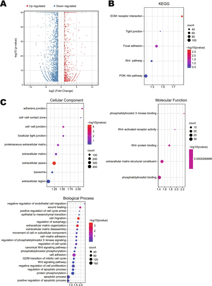
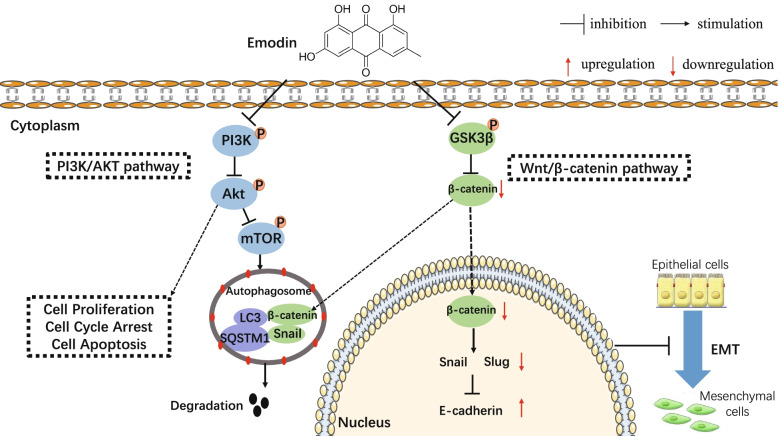
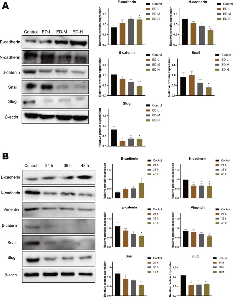
We found that emodin inhibited the growth of HepG2 cells in a dose- and time-dependent manner. In addition, emodin inhibited cell proliferation, induced S and G2/M phases arrest, and promoted apoptosis in HepG2 cells. The migration and invasion of HepG2 cells were also suppressed by emodin. Enrichment analysis revealed that DEGs involved in cell adhesion, cancer metastasis and cell cycle arrest. Moreover, western bolt results show that emodin-induced autophagy promotes Snail and β-catenin degradation. We also found that blocking autophagic flux after emodin treatment caused EMT reversal. Furthermore, the PI3K agonist Y-P 740 significantly reversed the phosphorylation levels of GSK3β and mTOR. These results indicated that emodin induced autophagy and inhibited the EMT in part through suppression of the PI3K/AKT/mTOR and Wnt/β-catenin pathways.
Our study indicated that emodin inhibited cell metastasis in HCC via the crosstalk between autophagy and EMT.
先前的研究表明,从大黄中提取的大黄素在多种人类癌症类型中具有抗增殖和抗转移作用。然而,大黄素在肝细胞癌(HCC)中的作用尚不清楚。
采用 EDU 和集落形成实验评估大黄素对增殖的影响。用划痕愈合实验评估大黄素处理后的 HCC 的迁移能力。Transwell 侵袭和迁移实验用于评估大黄素对 HCC 的抗迁移和抗侵袭作用。用 Annexin V-FITC/PI 分析凋亡。PI 染色用于分析细胞周期。采用 RNA 测序技术鉴定大黄素诱导的 HCC 中差异表达基因(DEGs)。用 mCherry-GFP-LC3 分析评估大黄素对 HepG2 细胞自噬流的影响。用 Western blot 检测上皮-间充质转化(EMT)、自噬、PI3K/AKT/mTOR 和 Wnt/β-catenin 信号通路的蛋白表达。
我们发现大黄素呈剂量和时间依赖性地抑制 HepG2 细胞的生长。此外,大黄素抑制 HepG2 细胞增殖,诱导 S 和 G2/M 期阻滞,并促进细胞凋亡。大黄素还抑制 HepG2 细胞的迁移和侵袭。富集分析显示,DEGs 涉及细胞黏附、癌症转移和细胞周期阻滞。此外,Western blot 结果表明,大黄素诱导的自噬促进了 Snail 和 β-catenin 的降解。我们还发现,大黄素处理后阻断自噬流导致 EMT 逆转。此外,PI3K 激动剂 Y-P 740 显著逆转了 GSK3β 和 mTOR 的磷酸化水平。这些结果表明,大黄素通过抑制 PI3K/AKT/mTOR 和 Wnt/β-catenin 通路诱导自噬并抑制 EMT。
本研究表明,大黄素通过自噬和 EMT 之间的相互作用抑制 HCC 细胞转移。