• 文献检索
  • 文档翻译
  • 深度研究
  • 学术资讯
  • Suppr Zotero 插件Zotero 插件
  • 邀请有礼
  • 套餐&价格
  • 历史记录
应用&插件
Suppr Zotero 插件Zotero 插件浏览器插件Mac 客户端Windows 客户端微信小程序
定价
高级版会员购买积分包购买API积分包
服务
文献检索文档翻译深度研究API 文档MCP 服务
关于我们
关于 Suppr公司介绍联系我们用户协议隐私条款
关注我们

Suppr 超能文献

核心技术专利:CN118964589B侵权必究
粤ICP备2023148730 号-1Suppr @ 2026

文献检索

告别复杂PubMed语法,用中文像聊天一样搜索,搜遍4000万医学文献。AI智能推荐,让科研检索更轻松。

立即免费搜索

文件翻译

保留排版,准确专业,支持PDF/Word/PPT等文件格式,支持 12+语言互译。

免费翻译文档

深度研究

AI帮你快速写综述,25分钟生成高质量综述,智能提取关键信息,辅助科研写作。

立即免费体验

颅底病变中感知错误的分析。

Analysis of perceptual errors in skull-base pathology.

作者信息

Vong Stephen, Chang Jennifer, Assadsangabi Reza, Ivanovic Vladimir

机构信息

Department of Radiology, UC Davis Health, Sacramento, CA, USA.

Department of Radiology, University of Southern California, Los Angeles, CA, USA.

出版信息

Neuroradiol J. 2023 Oct;36(5):515-523. doi: 10.1177/19714009221108679. Epub 2022 Jun 18.

DOI:10.1177/19714009221108679
PMID:35722674
原文链接:https://pmc.ncbi.nlm.nih.gov/articles/PMC10569193/
Abstract

PURPOSE

Medical errors result in significant mortality and morbidity. The purpose of this study is to analyze skull-base errors at a single tertiary institution, identify common anatomic sites of errors, and offer strategies to reduce errors in this region.

METHODS

A Neuroradiology Quality Assurance Database of radiologic errors was searched for attending physician computer tomography and magnetic resonance imaging errors in skull-base pathology from 2014 to 2020. Data were limited to CT and MRI reports. Errors were separated into four subcategories (tumor, trauma, vascular, and congenital) and further divided by relevant anatomic site.

RESULTS

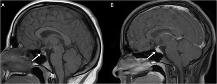
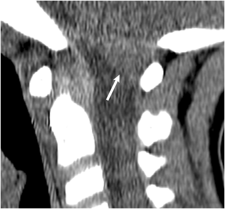
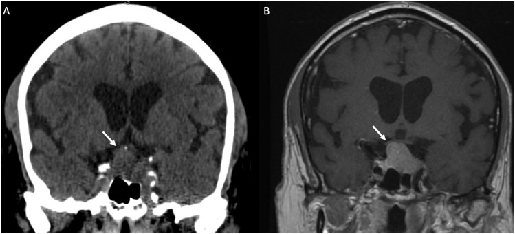
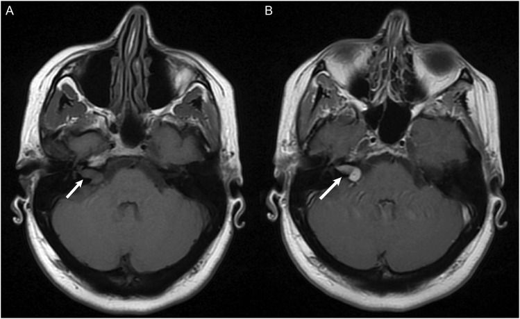
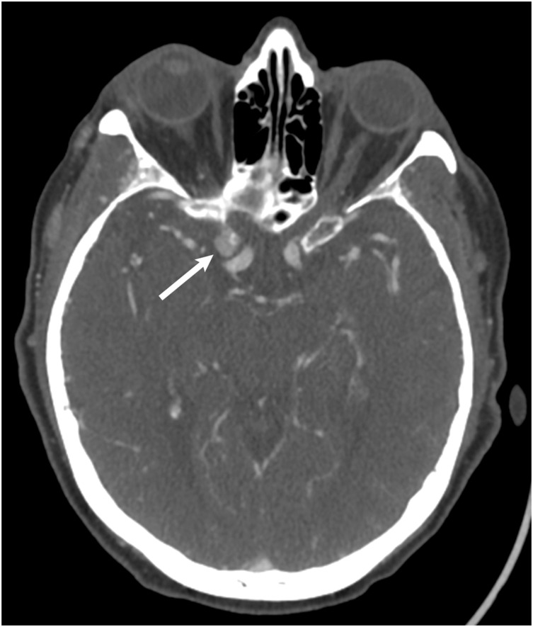
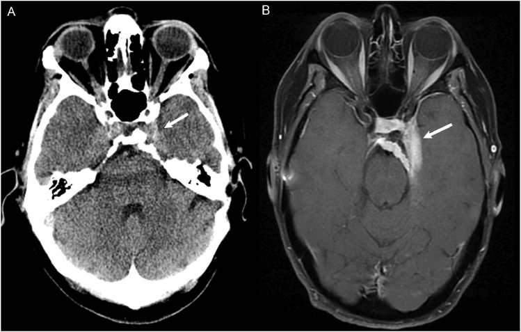
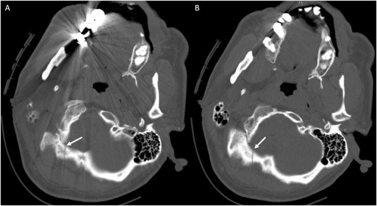
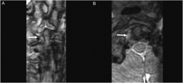
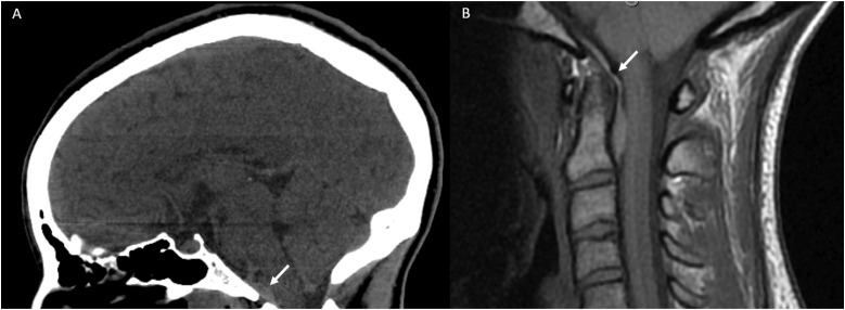
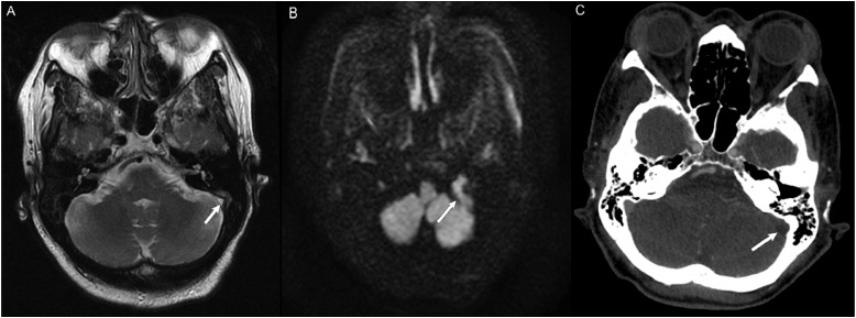
A total of 90 skull-based errors were identified. Most errors were perceptual (87%), with common study types including MRI Brain (39%) and CT Head (24%). Most common errors were tumors (55%), followed by trauma (24%), vascular (10%), and congenital (7%). Six anatomic sites were identified and encompassed over half of errors (58%): sella, occipital bone, cerebellopontine angle/internal auditory canal (CPA/IAC), foramen magnum and clivus, cavernous sinus, and dural venous sinus.

SUMMARY

Most of the skull-base errors were perceptual. Placing a strong emphasis on both the pathology and closely examining its critical anatomic site (sella, occipital bone, CPA/IAC, foramen magnum and clivus, cavernous sinus, and dural venous sinus) could potentially reduce up to 60% of errors in these regions.

摘要

目的

医疗差错会导致严重的死亡率和发病率。本研究的目的是分析一家三级医疗机构中的颅底差错,确定常见的解剖学差错部位,并提供减少该区域差错的策略。

方法

在一个神经放射学质量保证数据库中搜索2014年至2020年主治医生在颅底病变的计算机断层扫描和磁共振成像中的差错。数据仅限于CT和MRI报告。差错分为四个子类别(肿瘤、创伤、血管和先天性),并进一步按相关解剖部位划分。

结果

共识别出90例颅底差错。大多数差错是感知性的(87%),常见的检查类型包括脑部MRI(39%)和头部CT(24%)。最常见的差错是肿瘤(55%),其次是创伤(24%)、血管(10%)和先天性(7%)。确定了六个解剖部位,这些部位涵盖了一半以上的差错(58%):蝶鞍、枕骨、桥小脑角/内耳道(CPA/IAC)、枕大孔和斜坡、海绵窦和硬脑膜静脉窦。

总结

大多数颅底差错是感知性的。高度重视病变并仔细检查其关键解剖部位(蝶鞍、枕骨、CPA/IAC、枕大孔和斜坡、海绵窦和硬脑膜静脉窦)可能会减少这些区域高达60%的差错。

https://cdn.ncbi.nlm.nih.gov/pmc/blobs/f0db/10569193/5985a6a0c9b8/10.1177_19714009221108679-fig10.jpg
https://cdn.ncbi.nlm.nih.gov/pmc/blobs/f0db/10569193/7cee36eb0d4b/10.1177_19714009221108679-fig1.jpg
https://cdn.ncbi.nlm.nih.gov/pmc/blobs/f0db/10569193/8165550b612c/10.1177_19714009221108679-fig2.jpg
https://cdn.ncbi.nlm.nih.gov/pmc/blobs/f0db/10569193/7b28d9cc7245/10.1177_19714009221108679-fig3.jpg
https://cdn.ncbi.nlm.nih.gov/pmc/blobs/f0db/10569193/39d6a2218c83/10.1177_19714009221108679-fig4.jpg
https://cdn.ncbi.nlm.nih.gov/pmc/blobs/f0db/10569193/5eb01fda0d69/10.1177_19714009221108679-fig5.jpg
https://cdn.ncbi.nlm.nih.gov/pmc/blobs/f0db/10569193/fa2da1508c92/10.1177_19714009221108679-fig6.jpg
https://cdn.ncbi.nlm.nih.gov/pmc/blobs/f0db/10569193/34f7c99651ee/10.1177_19714009221108679-fig7.jpg
https://cdn.ncbi.nlm.nih.gov/pmc/blobs/f0db/10569193/8cfab58fead5/10.1177_19714009221108679-fig8.jpg
https://cdn.ncbi.nlm.nih.gov/pmc/blobs/f0db/10569193/f514cb9557fc/10.1177_19714009221108679-fig9.jpg
https://cdn.ncbi.nlm.nih.gov/pmc/blobs/f0db/10569193/5985a6a0c9b8/10.1177_19714009221108679-fig10.jpg

相似文献

1
Analysis of perceptual errors in skull-base pathology.颅底病变中感知错误的分析。
Neuroradiol J. 2023 Oct;36(5):515-523. doi: 10.1177/19714009221108679. Epub 2022 Jun 18.
2
Endoscopic transsphenoidal anterior petrosal approach for locally aggressive tumors involving the internal auditory canal, jugular fossa, and cavernous sinus.经鼻蝶窦前岩骨入路切除累及内听道、颈静脉窝和海绵窦的局部侵袭性肿瘤
J Neurosurg. 2017 Jan;126(1):212-221. doi: 10.3171/2016.1.JNS151979. Epub 2016 Apr 1.
3
Impact of Shift Volume on Neuroradiology Diagnostic Errors at a Large Tertiary Academic Center.大型三级学术中心轮班量对神经放射诊断错误的影响。
Acad Radiol. 2023 Aug;30(8):1584-1588. doi: 10.1016/j.acra.2022.08.035. Epub 2022 Sep 27.
4
Visualization of Dark Side of Skull Base with Surgical Navigation and Endoscopic Assistance: Extended Petrous Rhomboid and Rhomboid with Maxillary Nerve-Mandibular Nerve Vidian Corridor.手术导航和内镜辅助下颅底黑暗面的可视化:扩展的岩枕部和岩枕部,以及上颌神经-下颌神经翼管。
World Neurosurg. 2019 Sep;129:e134-e145. doi: 10.1016/j.wneu.2019.05.062. Epub 2019 May 17.
5
The incidence of invasion and metastasis of nasopharyngeal carcinoma at different anatomic sites in the skull base.颅底不同解剖部位鼻咽癌侵袭转移的发生率。
Anat Rec (Hoboken). 2012 Aug;295(8):1252-9. doi: 10.1002/ar.22507. Epub 2012 Jun 13.
6
Anatomy of the inferior petro-occipital vein and its relation to the base of the skull: application to surgical and endovascular procedures of the skull base.岩枕下静脉的解剖及其与颅底的关系:在颅底外科手术和血管内介入手术中的应用
Clin Anat. 2014 Jul;27(5):698-701. doi: 10.1002/ca.22268. Epub 2013 May 29.
7
Jugular bulb and skull base pathologies: proposal for a novel classification system for jugular bulb positions and microsurgical implications.颈静脉球和颅底病变:颈静脉球位置的新型分类系统及显微外科意义的建议。
Neurosurg Focus. 2018 Jul;45(1):E5. doi: 10.3171/2018.5.FOCUS18106.
8
Diagnostic Errors in Cerebrovascular Pathology: Retrospective Analysis of a Neuroradiology Database at a Large Tertiary Academic Medical Center.脑血管病病理学中的诊断错误:对大型三级学术医疗中心神经放射学数据库的回顾性分析。
AJNR Am J Neuroradiol. 2022 Sep;43(9):1271-1278. doi: 10.3174/ajnr.A7596. Epub 2022 Aug 4.
9
Analysis of misses in imaging of head and neck pathology by attending neuroradiologists at a single tertiary academic medical centre.单家三级学术医学中心神经放射科医师在头颈部病变影像学检查中的漏诊分析。
Clin Radiol. 2021 Oct;76(10):786.e9-786.e13. doi: 10.1016/j.crad.2021.06.011. Epub 2021 Jul 23.
10
Infraorbital nerve: a surgically relevant landmark for the pterygopalatine fossa, cavernous sinus, and anterolateral skull base in endoscopic transmaxillary approaches.眶下神经:经上颌窦内镜入路中翼腭窝、海绵窦和颅前窝外侧底的手术相关解剖标志
J Neurosurg. 2016 Dec;125(6):1460-1468. doi: 10.3171/2015.9.JNS151099. Epub 2016 Mar 4.

引用本文的文献

1
Shift Volume Directly Impacts Neuroradiology Error Rate at a Large Academic Medical Center: The Case for Volume Limits.大容量直接影响大型学术医疗中心的神经放射学错误率:限制容量的案例。
AJNR Am J Neuroradiol. 2024 Apr 8;45(4):374-378. doi: 10.3174/ajnr.A8119.
2
Peer Learning in Neuroradiology: Not as Easy as It Sounds.神经放射学中的同伴学习:并非听起来那么容易。
AJNR Am J Neuroradiol. 2023 Oct;44(10):1109-1115. doi: 10.3174/ajnr.A7973.

本文引用的文献

1
Sellar, suprasellar, and parasellar masses: Imaging features and neurosurgical approaches.鞍区、鞍上和鞍旁肿块:影像学特征和神经外科入路。
Neuroradiol J. 2022 Jun;35(3):269-283. doi: 10.1177/19714009211055195. Epub 2021 Dec 2.
2
Analysis of misses in imaging of head and neck pathology by attending neuroradiologists at a single tertiary academic medical centre.单家三级学术医学中心神经放射科医师在头颈部病变影像学检查中的漏诊分析。
Clin Radiol. 2021 Oct;76(10):786.e9-786.e13. doi: 10.1016/j.crad.2021.06.011. Epub 2021 Jul 23.
3
Unilateral Nonvisualization of a Transverse Dural Sinus on Phase-Contrast MRV: Frequency and Differentiation from Sinus Thrombosis on Noncontrast MRI.
磁共振静脉血管成像相位对比技术中单侧横窦未显影:在非对比 MRI 上与窦血栓形成的鉴别诊断及发生率。
AJNR Am J Neuroradiol. 2020 Jan;41(1):115-121. doi: 10.3174/ajnr.A6337.
4
Risk Factors for Perceptual-versus-Interpretative Errors in Diagnostic Neuroradiology.诊断神经放射学中感知与解释错误的风险因素。
AJNR Am J Neuroradiol. 2019 Aug;40(8):1252-1256. doi: 10.3174/ajnr.A6125. Epub 2019 Jul 11.
5
Fundamentals of Diagnostic Error in Imaging.医学影像学诊断错误基础
Radiographics. 2018 Oct;38(6):1845-1865. doi: 10.1148/rg.2018180021.
6
Radiologic errors, past, present and future.放射学错误,过去、现在与未来。
Diagnosis (Berl). 2014 Jan 1;1(1):79-84. doi: 10.1515/dx-2013-0012.
7
Heuristics and Cognitive Error in Medical Imaging.医学影像学中的启发式和认知错误。
AJR Am J Roentgenol. 2018 May;210(5):1097-1105. doi: 10.2214/AJR.17.18907. Epub 2018 Mar 12.
8
Bias in Radiology: The How and Why of Misses and Misinterpretations.放射学中的偏倚:漏诊和误诊的原因与方式。
Radiographics. 2018 Jan-Feb;38(1):236-247. doi: 10.1148/rg.2018170107. Epub 2017 Dec 1.
9
Effect of Shift, Schedule, and Volume on Interpretive Accuracy: A Retrospective Analysis of 2.9 Million Radiologic Examinations.班次、轮班时间和工作量对诊断准确性的影响:290 万次放射检查的回顾性分析。
Radiology. 2018 Apr;287(1):205-212. doi: 10.1148/radiol.2017170555. Epub 2017 Nov 20.
10
Trainees May Add Value to Patient Care by Decreasing Addendum Utilization in Radiology Reports.实习生可通过减少放射学报告中的补遗使用为患者护理增添价值。
AJR Am J Roentgenol. 2017 Nov;209(5):976-981. doi: 10.2214/AJR.17.18339. Epub 2017 Aug 4.