• 文献检索
  • 文档翻译
  • 深度研究
  • 学术资讯
  • Suppr Zotero 插件Zotero 插件
  • 邀请有礼
  • 套餐&价格
  • 历史记录
应用&插件
Suppr Zotero 插件Zotero 插件浏览器插件Mac 客户端Windows 客户端微信小程序
定价
高级版会员购买积分包购买API积分包
服务
文献检索文档翻译深度研究API 文档MCP 服务
关于我们
关于 Suppr公司介绍联系我们用户协议隐私条款
关注我们

Suppr 超能文献

核心技术专利:CN118964589B侵权必究
粤ICP备2023148730 号-1Suppr @ 2026

文献检索

告别复杂PubMed语法,用中文像聊天一样搜索,搜遍4000万医学文献。AI智能推荐,让科研检索更轻松。

立即免费搜索

文件翻译

保留排版,准确专业,支持PDF/Word/PPT等文件格式,支持 12+语言互译。

免费翻译文档

深度研究

AI帮你快速写综述,25分钟生成高质量综述,智能提取关键信息,辅助科研写作。

立即免费体验

青光眼结构-功能建模中机器学习方法的比较。

Comparison of machine learning approaches for structure-function modeling in glaucoma.

机构信息

SERI-NTU Advanced Ocular Engineering (STANCE), Singapore.

School of Chemical and Biomedical Engineering, Nanyang Technological University, Singapore.

出版信息

Ann N Y Acad Sci. 2022 Sep;1515(1):237-248. doi: 10.1111/nyas.14844. Epub 2022 Jun 21.

DOI:10.1111/nyas.14844
PMID:35729796
原文链接:https://pmc.ncbi.nlm.nih.gov/articles/PMC10946805/
Abstract

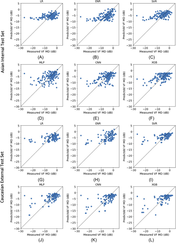
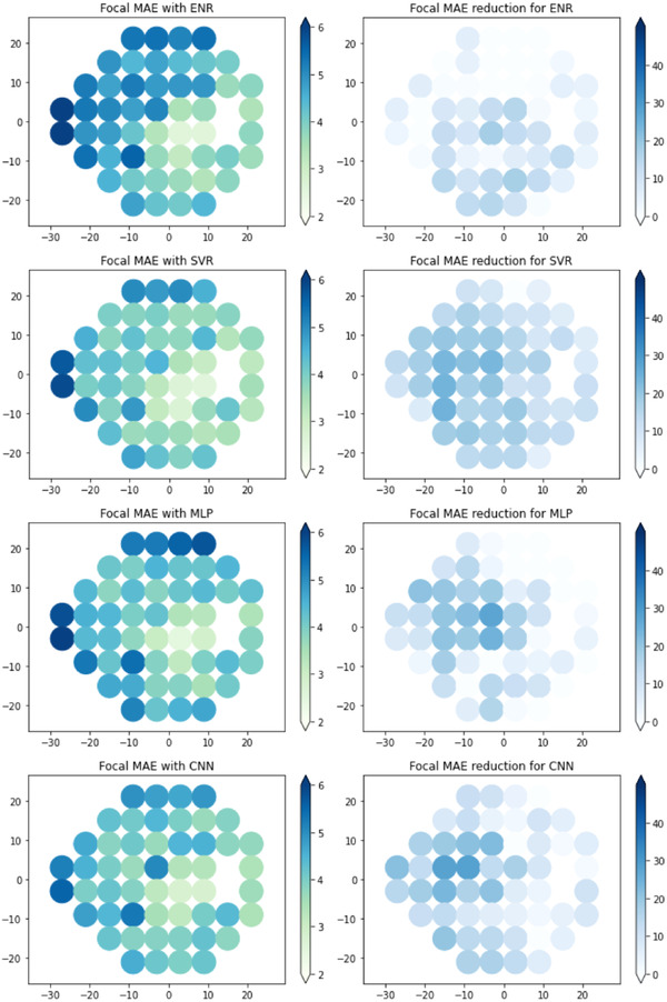
To evaluate machine learning (ML) approaches for structure-function modeling to estimate visual field (VF) loss in glaucoma, models from different ML approaches were trained on optical coherence tomography thickness measurements to estimate global VF mean deviation (VF MD) and focal VF loss from 24-2 standard automated perimetry. The models were compared using mean absolute errors (MAEs). Baseline MAEs were obtained from the VF values and their means. Data of 832 eyes from 569 participants were included, with 537 Asian eyes for training, and 148 Asian and 111 Caucasian eyes set aside as the respective test sets. All ML models performed significantly better than baseline. Gradient-boosted trees (XGB) achieved the lowest MAE of 3.01 (95% CI: 2.57, 3.48) dB and 3.04 (95% CI: 2.59, 3.99) dB for VF MD estimation in the Asian and Caucasian test sets, although difference between models was not significant. In focal VF estimation, XGB achieved median MAEs of 4.44 [IQR 3.45-5.17] dB and 3.87 [IQR 3.64-4.22] dB across the 24-2 VF for the Asian and Caucasian test sets and was comparable to VF estimates from support vector regression (SVR) models. VF estimates from both XGB and SVR were significantly better than the other models. These results show that XGB and SVR could potentially be used for both global and focal structure-function modeling in glaucoma.

摘要

为了评估机器学习(ML)方法在评估青光眼视野(VF)损失中的结构-功能建模,从不同的 ML 方法中训练模型,使用光学相干断层扫描(OCT)厚度测量来估计全球 VF 平均偏差(VF MD)和从 24-2 标准自动视野计获得的焦点 VF 损失。通过平均绝对误差(MAE)来比较模型。基线 MAE 是从 VF 值及其平均值中获得的。纳入了 569 名参与者的 832 只眼的数据,其中 537 只为亚洲眼,用于训练,148 只为亚洲眼和 111 只为高加索眼分别作为各自的测试集。所有 ML 模型的性能均明显优于基线。梯度提升树(XGB)在亚洲和高加索测试集中,VF MD 估计的 MAE 分别达到 3.01(95%CI:2.57,3.48)dB 和 3.04(95%CI:2.59,3.99)dB,达到最低水平,尽管模型之间的差异不显著。在焦点 VF 估计中,XGB 在亚洲和高加索测试集中分别达到 24-2 VF 的中位数 MAE 为 4.44[IQR 3.45-5.17]dB 和 3.87[IQR 3.64-4.22]dB,与支持向量回归(SVR)模型的 VF 估计相当。XGB 和 SVR 的 VF 估计均明显优于其他模型。这些结果表明,XGB 和 SVR 可能可用于青光眼的全局和焦点结构-功能建模。

https://cdn.ncbi.nlm.nih.gov/pmc/blobs/7f5c/10946805/e35773a08cdb/NYAS-1515-237-g001.jpg
https://cdn.ncbi.nlm.nih.gov/pmc/blobs/7f5c/10946805/b42d054520a2/NYAS-1515-237-g005.jpg
https://cdn.ncbi.nlm.nih.gov/pmc/blobs/7f5c/10946805/3c61d536506f/NYAS-1515-237-g002.jpg
https://cdn.ncbi.nlm.nih.gov/pmc/blobs/7f5c/10946805/d39de589530c/NYAS-1515-237-g003.jpg
https://cdn.ncbi.nlm.nih.gov/pmc/blobs/7f5c/10946805/e35773a08cdb/NYAS-1515-237-g001.jpg

相似文献

1
Comparison of machine learning approaches for structure-function modeling in glaucoma.青光眼结构-功能建模中机器学习方法的比较。
Ann N Y Acad Sci. 2022 Sep;1515(1):237-248. doi: 10.1111/nyas.14844. Epub 2022 Jun 21.
2
Deep Learning Estimation of 10-2 and 24-2 Visual Field Metrics Based on Thickness Maps from Macula OCT.基于黄斑 OCT 厚度图的深度学习估算 10-2 和 24-2 视野指标。
Ophthalmology. 2021 Nov;128(11):1534-1548. doi: 10.1016/j.ophtha.2021.04.022. Epub 2021 Apr 23.
3
Deep Learning Estimation of 10-2 Visual Field Map Based on Macular Optical Coherence Tomography Angiography Measurements.基于黄斑光学相干断层扫描血管造影测量的10-2视野图的深度学习估计
Am J Ophthalmol. 2024 Jan;257:187-200. doi: 10.1016/j.ajo.2023.09.014. Epub 2023 Sep 19.
4
Pointwise Visual Field Estimation From Optical Coherence Tomography in Glaucoma Using Deep Learning.基于深度学习的青光眼光学相干断层扫描的逐点视野估计。
Transl Vis Sci Technol. 2022 Aug 1;11(8):22. doi: 10.1167/tvst.11.8.22.
5
Deep learning model to predict visual field in central 10° from optical coherence tomography measurement in glaucoma.深度学习模型预测青光眼光学相干断层扫描测量的中央 10°视野。
Br J Ophthalmol. 2021 Apr;105(4):507-513. doi: 10.1136/bjophthalmol-2019-315600. Epub 2020 Jun 27.
6
Combining OCT and OCTA for Focal Structure-Function Modeling in Early Primary Open-Angle Glaucoma.将 OCT 和 OCTA 相结合进行早期原发性开角型青光眼的焦点结构-功能建模。
Invest Ophthalmol Vis Sci. 2021 Dec 1;62(15):8. doi: 10.1167/iovs.62.15.8.
7
Comparative study of retinal nerve fiber layer measurement by StratusOCT and GDx VCC, II: structure/function regression analysis in glaucoma.StratusOCT与GDx VCC测量视网膜神经纤维层的对比研究,II:青光眼的结构/功能回归分析
Invest Ophthalmol Vis Sci. 2005 Oct;46(10):3702-11. doi: 10.1167/iovs.05-0490.
8
Effect of focal lamina cribrosa defect on glaucomatous visual field progression.视盘局限性筛板缺损对青光眼视野进展的影响。
Ophthalmology. 2014 Aug;121(8):1524-30. doi: 10.1016/j.ophtha.2014.02.017. Epub 2014 Mar 31.
9
Factors affecting rates of visual field progression in glaucoma patients with optic disc hemorrhage.影响伴有视盘出血的青光眼患者视野进展速度的因素。
Ophthalmology. 2010 Jan;117(1):24-9. doi: 10.1016/j.ophtha.2009.06.028. Epub 2009 Nov 6.
10
Relationship Between Central Retinal Vessel Trunk Location and Visual Field Loss in Glaucoma.青光眼患者视网膜中央血管主干位置与视野缺损的关系
Am J Ophthalmol. 2017 Apr;176:53-60. doi: 10.1016/j.ajo.2017.01.001. Epub 2017 Jan 11.

引用本文的文献

1
Interpretable longitudinal glaucoma visual field estimation deep learning system from fundus images and clinical narratives.基于眼底图像和临床记录的可解释性纵向青光眼视野估计深度学习系统
NPJ Digit Med. 2025 Jul 1;8(1):389. doi: 10.1038/s41746-025-01750-8.
2
Automated learning of glaucomatous visual fields from OCT images using a comprehensive, segmentation-free 3D convolutional neural network model.使用全面的、无需分割的3D卷积神经网络模型从光学相干断层扫描(OCT)图像中自动学习青光眼视野
Sci Rep. 2025 Apr 18;15(1):13395. doi: 10.1038/s41598-025-98511-0.
3
Detecting glaucoma worsening using optical coherence tomography derived visual field estimates.

本文引用的文献

1
Pointwise Visual Field Estimation From Optical Coherence Tomography in Glaucoma Using Deep Learning.基于深度学习的青光眼光学相干断层扫描的逐点视野估计。
Transl Vis Sci Technol. 2022 Aug 1;11(8):22. doi: 10.1167/tvst.11.8.22.
2
Policy-Driven, Multimodal Deep Learning for Predicting Visual Fields from the Optic Disc and OCT Imaging.基于策略驱动的多模态深度学习,用于从视盘和光学相干断层扫描(OCT)成像预测视野。
Ophthalmology. 2022 Jul;129(7):781-791. doi: 10.1016/j.ophtha.2022.02.017. Epub 2022 Feb 21.
3
Combining vascular and nerve fiber layer thickness measurements to model glaucomatous focal visual field loss.
使用光学相干断层扫描得出的视野估计来检测青光眼病情恶化。
Sci Rep. 2025 Feb 11;15(1):5013. doi: 10.1038/s41598-025-86217-2.
4
Artificial intelligence in glaucoma: opportunities, challenges, and future directions.人工智能在青光眼领域的应用:机遇、挑战与未来方向。
Biomed Eng Online. 2023 Dec 16;22(1):126. doi: 10.1186/s12938-023-01187-8.
5
Pointwise Visual Field Estimation From Optical Coherence Tomography in Glaucoma Using Deep Learning.基于深度学习的青光眼光学相干断层扫描的逐点视野估计。
Transl Vis Sci Technol. 2022 Aug 1;11(8):22. doi: 10.1167/tvst.11.8.22.
将血管和神经纤维层厚度测量相结合,以模拟青光眼的局灶性视野损失。
Ann N Y Acad Sci. 2022 May;1511(1):133-141. doi: 10.1111/nyas.14732. Epub 2022 Jan 14.
4
Predicting Visual Fields From Optical Coherence Tomography via an Ensemble of Deep Representation Learners.基于深度表示学习器集成的光学相干断层扫描预测视野。
Am J Ophthalmol. 2022 Jun;238:52-65. doi: 10.1016/j.ajo.2021.12.020. Epub 2022 Jan 5.
5
Combining OCT and OCTA for Focal Structure-Function Modeling in Early Primary Open-Angle Glaucoma.将 OCT 和 OCTA 相结合进行早期原发性开角型青光眼的焦点结构-功能建模。
Invest Ophthalmol Vis Sci. 2021 Dec 1;62(15):8. doi: 10.1167/iovs.62.15.8.
6
Estimating the Severity of Visual Field Damage From Retinal Nerve Fiber Layer Thickness Measurements With Artificial Intelligence.利用人工智能估算视网膜神经纤维层厚度测量的视野损伤严重程度。
Transl Vis Sci Technol. 2021 Aug 2;10(9):16. doi: 10.1167/tvst.10.9.16.
7
Segregation of neuronal-vascular components in a retinal nerve fiber layer for thickness measurement using OCT and OCT angiography.利用光学相干断层扫描(OCT)和OCT血管造影术测量视网膜神经纤维层厚度时神经元-血管成分的分离
Biomed Opt Express. 2021 May 7;12(6):3228-3240. doi: 10.1364/BOE.420507. eCollection 2021 Jun 1.
8
Measuring optic nerve head perfusion to monitor glaucoma: a study on structure-function relationships using laser speckle flowgraphy.测量视神经乳头灌注以监测青光眼:一项使用激光散斑血流图研究结构-功能关系的研究
Acta Ophthalmol. 2022 Feb;100(1):e181-e191. doi: 10.1111/aos.14862. Epub 2021 Apr 20.
9
Focal Structure-Function Relationships in Primary Open-Angle Glaucoma Using OCT and OCT-A Measurements.应用 OCT 和 OCT-A 测量评估原发性开角型青光眼的局灶结构-功能关系。
Invest Ophthalmol Vis Sci. 2020 Dec 1;61(14):33. doi: 10.1167/iovs.61.14.33.
10
Estimating Global Visual Field Indices in Glaucoma by Combining Macula and Optic Disc OCT Scans Using 3-Dimensional Convolutional Neural Networks.利用三维卷积神经网络结合黄斑和视盘 OCT 扫描估算青光眼的全球视野指数。
Ophthalmol Glaucoma. 2021 Jan-Feb;4(1):102-112. doi: 10.1016/j.ogla.2020.07.002. Epub 2020 Jul 11.