• 文献检索
  • 文档翻译
  • 深度研究
  • 学术资讯
  • Suppr Zotero 插件Zotero 插件
  • 邀请有礼
  • 套餐&价格
  • 历史记录
应用&插件
Suppr Zotero 插件Zotero 插件浏览器插件Mac 客户端Windows 客户端微信小程序
定价
高级版会员购买积分包购买API积分包
服务
文献检索文档翻译深度研究API 文档MCP 服务
关于我们
关于 Suppr公司介绍联系我们用户协议隐私条款
关注我们

Suppr 超能文献

核心技术专利:CN118964589B侵权必究
粤ICP备2023148730 号-1Suppr @ 2026

文献检索

告别复杂PubMed语法,用中文像聊天一样搜索,搜遍4000万医学文献。AI智能推荐,让科研检索更轻松。

立即免费搜索

文件翻译

保留排版,准确专业,支持PDF/Word/PPT等文件格式,支持 12+语言互译。

免费翻译文档

深度研究

AI帮你快速写综述,25分钟生成高质量综述,智能提取关键信息,辅助科研写作。

立即免费体验

结合实时透射电子显微镜和集成机器学习-粒子滤波器估计的纳米尺度缺陷评估框架

Nanoscale defect evaluation framework combining real-time transmission electron microscopy and integrated machine learning-particle filter estimation.

作者信息

Sasaki K, Muramatsu M, Hirayama K, Endo K, Murayama M

机构信息

Department of Science for Open and Environmental Systems, Graduate School of Keio University, 3-14-1, Hiyoshi, Kohoku-ku, Kanagawa, 233-8522, Japan.

Department of Materials Science and Engineering, Virginia Tech, Blacksburg, VA, 24061, USA.

出版信息

Sci Rep. 2022 Jun 22;12(1):10525. doi: 10.1038/s41598-022-13878-8.

DOI:10.1038/s41598-022-13878-8
PMID:35732650
原文链接:https://pmc.ncbi.nlm.nih.gov/articles/PMC9217921/
Abstract

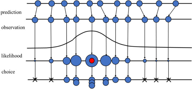
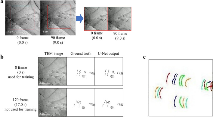
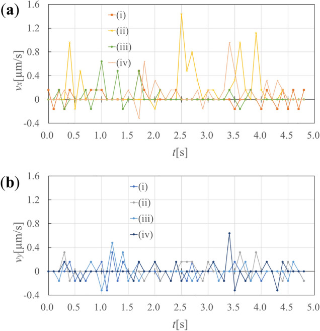
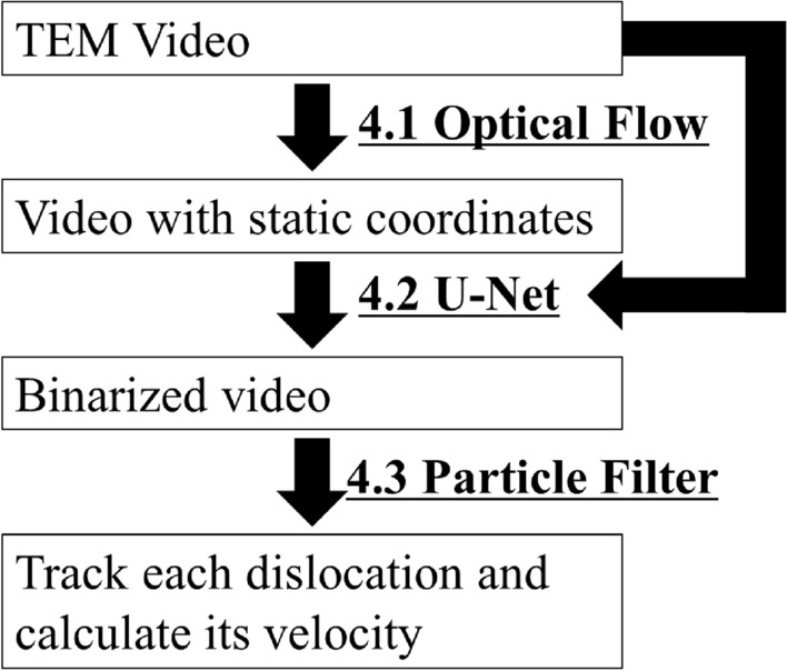
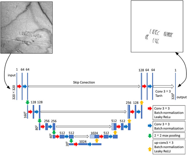
Observation of dynamic processes by transmission electron microscopy (TEM) is an attractive technique to experimentally analyze materials' nanoscale phenomena and understand the microstructure-properties relationships in nanoscale. Even if spatial and temporal resolutions of real-time TEM increase significantly, it is still difficult to say that the researchers quantitatively evaluate the dynamic behavior of defects. Images in TEM video are a two-dimensional projection of three-dimensional space phenomena, thus missing information must be existed that makes image's uniquely accurate interpretation challenging. Therefore, even though they are still a clustering high-dimensional data and can be compressed to two-dimensional, conventional statistical methods for analyzing images may not be powerful enough to track nanoscale behavior by removing various artifacts associated with experiment; and automated and unbiased processing tools for such big-data are becoming mission-critical to discover knowledge about unforeseen behavior. We have developed a method to quantitative image analysis framework to resolve these problems, in which machine learning and particle filter estimation are uniquely combined. The quantitative and automated measurement of the dislocation velocity in an Fe-31Mn-3Al-3Si autunitic steel subjected to the tensile deformation was performed to validate the framework, and an intermittent motion of the dislocations was quantitatively analyzed. The framework is successfully classifying, identifying and tracking nanoscale objects; these are not able to be accurately implemented by the conventional mean-path based analysis.

摘要

通过透射电子显微镜(TEM)观察动态过程是一种颇具吸引力的技术,可用于实验分析材料的纳米级现象,并理解纳米级的微观结构与性能之间的关系。即便实时TEM的空间和时间分辨率显著提高,但要说研究人员能够定量评估缺陷的动态行为仍非易事。TEM视频中的图像是三维空间现象的二维投影,因此必然存在缺失信息,这使得对图像进行唯一准确的解释颇具挑战性。所以,尽管它们仍是聚类的高维数据且可压缩为二维,但用于分析图像的传统统计方法可能不足以通过去除与实验相关的各种伪像来追踪纳米级行为;而针对此类大数据的自动化且无偏差的处理工具对于发现意外行为的知识正变得至关重要。我们开发了一种定量图像分析框架方法来解决这些问题,其中机器学习和粒子滤波器估计被独特地结合在一起。对经拉伸变形的Fe-31Mn-3Al-3Si奥氏体钢中的位错速度进行了定量和自动化测量,以验证该框架,并对位错的间歇运动进行了定量分析。该框架成功地对纳米级物体进行了分类、识别和跟踪;而这些通过传统的基于平均路径的分析无法准确实现。

https://cdn.ncbi.nlm.nih.gov/pmc/blobs/9b9f/9217921/ccb3efb7b6ac/41598_2022_13878_Fig8_HTML.jpg
https://cdn.ncbi.nlm.nih.gov/pmc/blobs/9b9f/9217921/1355ad22c111/41598_2022_13878_Fig1_HTML.jpg
https://cdn.ncbi.nlm.nih.gov/pmc/blobs/9b9f/9217921/e2c0a0561e9f/41598_2022_13878_Fig2_HTML.jpg
https://cdn.ncbi.nlm.nih.gov/pmc/blobs/9b9f/9217921/89643ade7b71/41598_2022_13878_Fig3_HTML.jpg
https://cdn.ncbi.nlm.nih.gov/pmc/blobs/9b9f/9217921/01e04e2ec09b/41598_2022_13878_Fig4_HTML.jpg
https://cdn.ncbi.nlm.nih.gov/pmc/blobs/9b9f/9217921/dbf5ebc283ab/41598_2022_13878_Fig5_HTML.jpg
https://cdn.ncbi.nlm.nih.gov/pmc/blobs/9b9f/9217921/3b48199d89f1/41598_2022_13878_Fig6_HTML.jpg
https://cdn.ncbi.nlm.nih.gov/pmc/blobs/9b9f/9217921/f8ea4f3c54b1/41598_2022_13878_Fig7_HTML.jpg
https://cdn.ncbi.nlm.nih.gov/pmc/blobs/9b9f/9217921/ccb3efb7b6ac/41598_2022_13878_Fig8_HTML.jpg

相似文献

1
Nanoscale defect evaluation framework combining real-time transmission electron microscopy and integrated machine learning-particle filter estimation.结合实时透射电子显微镜和集成机器学习-粒子滤波器估计的纳米尺度缺陷评估框架
Sci Rep. 2022 Jun 22;12(1):10525. doi: 10.1038/s41598-022-13878-8.
2
In situ transmission electron microscopy observation of dislocation motion in 9Cr steel at elevated temperatures: influence of shear stress on dislocation behavior.9Cr钢在高温下位错运动的原位透射电子显微镜观察:剪切应力对位错行为的影响。
Microscopy (Oxf). 2014 Dec;63(6):449-61. doi: 10.1093/jmicro/dfu035. Epub 2014 Oct 8.
3
Deep Learning Segmentation of Complex Features in Atomic-Resolution Phase-Contrast Transmission Electron Microscopy Images.原子分辨率相衬透射电子显微镜图像中复杂特征的深度学习分割
Microsc Microanal. 2021 Aug;27(4):804-814. doi: 10.1017/S1431927621000167.
4
Prior data assisted compressed sensing: a novel MR imaging strategy for real time tracking of lung tumors.先前数据辅助压缩感知:一种用于实时追踪肺部肿瘤的新型磁共振成像策略。
Med Phys. 2014 Aug;41(8):082301. doi: 10.1118/1.4885960.
5
Deformation mechanisms in free-standing nanoscale thin films: a quantitative in situ transmission electron microscope study.独立纳米级薄膜中的变形机制:定量原位透射电子显微镜研究。
Proc Natl Acad Sci U S A. 2004 Apr 27;101(17):6335-40. doi: 10.1073/pnas.0400066101. Epub 2004 Apr 14.
6
Identification of sampling patterns for high-resolution compressed sensing MRI of porous materials: 'learning' from X-ray microcomputed tomography data.多孔材料高分辨率压缩感知 MRI 采样模式的识别:从 X 射线微计算机断层扫描数据中“学习”。
J Microsc. 2019 Nov;276(2):63-81. doi: 10.1111/jmi.12837. Epub 2019 Nov 6.
7
3D cine-magnetic resonance imaging using spatial and temporal implicit neural representation learning (STINR-MR).使用空间和时间隐式神经表示学习的3D电影磁共振成像(STINR-MR)。
Phys Med Biol. 2024 Apr 15;69(9):095007. doi: 10.1088/1361-6560/ad33b7.
8
Creating ground truth for nanocrystal morphology: a fully automated pipeline for unbiased transmission electron microscopy analysis.创建纳米晶体形态的真实数据:一种用于无偏倚透射电子显微镜分析的全自动流程。
Nanoscale. 2022 Oct 27;14(41):15327-15339. doi: 10.1039/d2nr04292d.
9
Statistical Characterization of the Morphologies of Nanoparticles through Machine Learning Based Electron Microscopy Image Analysis.通过基于机器学习的电子显微镜图像分析对纳米颗粒形态进行统计表征。
ACS Nano. 2020 Dec 22;14(12):17125-17133. doi: 10.1021/acsnano.0c06809. Epub 2020 Nov 24.
10
Machine Learning to Reveal Nanoparticle Dynamics from Liquid-Phase TEM Videos.利用机器学习从液相透射电子显微镜视频中揭示纳米颗粒动力学
ACS Cent Sci. 2020 Aug 26;6(8):1421-1430. doi: 10.1021/acscentsci.0c00430. Epub 2020 Jul 6.

引用本文的文献

1
Machine-learning-aided analysis of relationship between crystal defects and macroscopic mechanical properties of TWIP steel.基于机器学习辅助分析TWIP钢晶体缺陷与宏观力学性能之间的关系
Sci Rep. 2025 Apr 25;15(1):14435. doi: 10.1038/s41598-025-99319-8.

本文引用的文献

1
Predicting material microstructure evolution via data-driven machine learning.通过数据驱动的机器学习预测材料微观结构演变。
Patterns (N Y). 2021 Jun 18;2(7):100285. doi: 10.1016/j.patter.2021.100285. eCollection 2021 Jul 9.
2
A deep learning approach to identifying immunogold particles in electron microscopy images.一种用于在电子显微镜图像中识别免疫金颗粒的深度学习方法。
Sci Rep. 2021 Apr 8;11(1):7771. doi: 10.1038/s41598-021-87015-2.
3
Towards data-driven next-generation transmission electron microscopy.迈向数据驱动的下一代透射电子显微镜。
Nat Mater. 2021 Mar;20(3):274-279. doi: 10.1038/s41563-020-00833-z.
4
Sparse Scanning Electron Microscopy Data Acquisition and Deep Neural Networks for Automated Segmentation in Connectomics.稀疏扫描电子显微镜数据采集和深度学习神经网络在连接组学中的自动分割。
Microsc Microanal. 2020 Jun;26(3):403-412. doi: 10.1017/S1431927620001361.
5
Deep Learning for Semantic Segmentation of Defects in Advanced STEM Images of Steels.深度学习在钢铁高级 STEM 图像缺陷的语义分割中的应用。
Sci Rep. 2019 Sep 4;9(1):12744. doi: 10.1038/s41598-019-49105-0.
6
Machine learning plastic deformation of crystals.机器学习在晶体变形中的应用。
Nat Commun. 2018 Dec 13;9(1):5307. doi: 10.1038/s41467-018-07737-2.
7
Trainable Weka Segmentation: a machine learning tool for microscopy pixel classification.可训练的 WEKA 分割:一种用于显微镜像素分类的机器学习工具。
Bioinformatics. 2017 Aug 1;33(15):2424-2426. doi: 10.1093/bioinformatics/btx180.
8
SegNet: A Deep Convolutional Encoder-Decoder Architecture for Image Segmentation.SegNet:一种用于图像分割的深度卷积编解码器架构。
IEEE Trans Pattern Anal Mach Intell. 2017 Dec;39(12):2481-2495. doi: 10.1109/TPAMI.2016.2644615. Epub 2017 Jan 2.
9
Fully Convolutional Networks for Semantic Segmentation.全卷积网络用于语义分割。
IEEE Trans Pattern Anal Mach Intell. 2017 Apr;39(4):640-651. doi: 10.1109/TPAMI.2016.2572683. Epub 2016 May 24.
10
A predictive machine learning approach for microstructure optimization and materials design.一种用于微观结构优化和材料设计的预测性机器学习方法。
Sci Rep. 2015 Jun 23;5:11551. doi: 10.1038/srep11551.