Department of Radiotherapy, ICANS, Institut Cancérologie Strasbourg Europe, 17 Rue Albert Calmette, 67200, Strasbourg Cedex, France.
Department of Radiotherapy, Institut Gustave Roussy, Paris-Saclay University, Villejuif, France.
Sci Rep. 2022 Jun 22;12(1):10502. doi: 10.1038/s41598-022-13739-4.
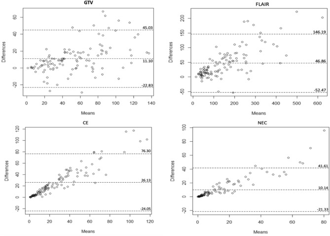
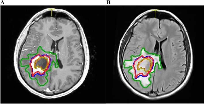
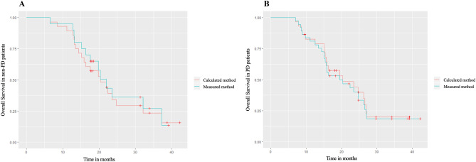
In glioblastoma, the response to treatment assessment is essentially based on the 2D tumor size evolution but remains disputable. Volumetric approaches were evaluated for a more accurate estimation of tumor size. This study included 57 patients and compared two volume measurement methods to determine the size of different glioblastoma regions of interest: the contrast-enhancing area, the necrotic area, the gross target volume and the volume of the edema area. The two methods, the ellipsoid formula (the calculated method) and the manual delineation (the measured method) showed a high correlation to determine glioblastoma volume and a high agreement to classify patients assessment response to treatment according to RANO criteria. This study revealed that calculated and measured methods could be used in clinical practice to estimate glioblastoma volume size and to evaluate tumor size evolution.
在胶质母细胞瘤中,治疗反应评估主要基于 2D 肿瘤大小的演变,但仍然存在争议。已经评估了各种体积评估方法,以更准确地估计肿瘤大小。本研究纳入了 57 名患者,比较了两种体积测量方法,以确定不同胶质母细胞瘤感兴趣区域的大小:增强区域、坏死区域、大体肿瘤靶区和水肿区域体积。这两种方法,即椭球公式(计算法)和手动勾画(测量法),显示出高度相关性,可用于确定胶质母细胞瘤的体积,并且高度一致,可以根据 RANO 标准对患者的治疗反应进行分类。本研究表明,计算法和测量法可在临床实践中用于估计胶质母细胞瘤的体积大小,并评估肿瘤大小的演变。