Graduate School, Kunming Medical University, Kunming, Yunnan, China.
Department of Orthopaedics, People's Liberation Army Joint Logistic Support Force 920th Hospital, Kunming, Yunnan, China.
Bioengineered. 2022 Jun;13(6):14270-14281. doi: 10.1080/21655979.2022.2085560.
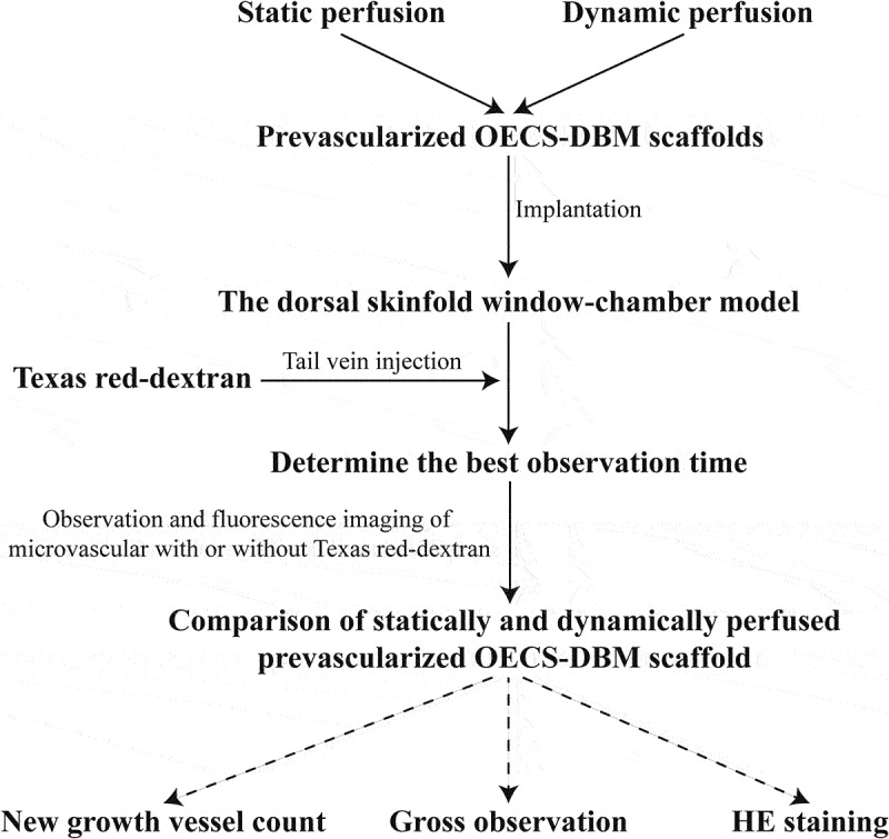
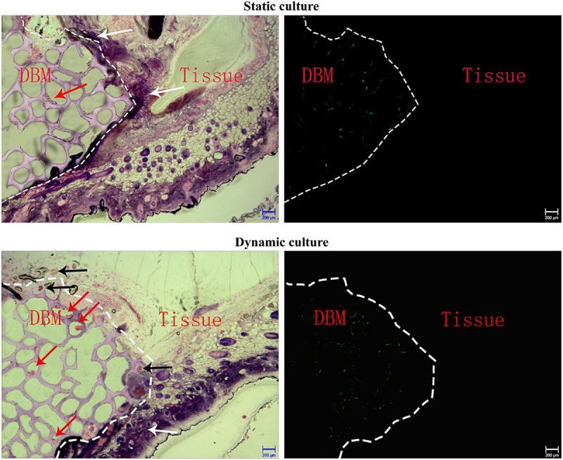
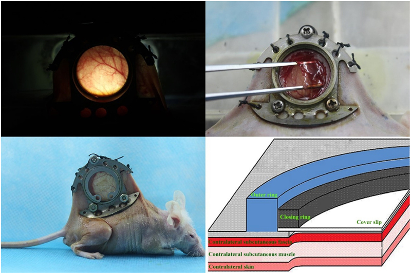
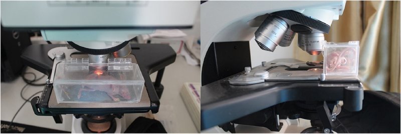
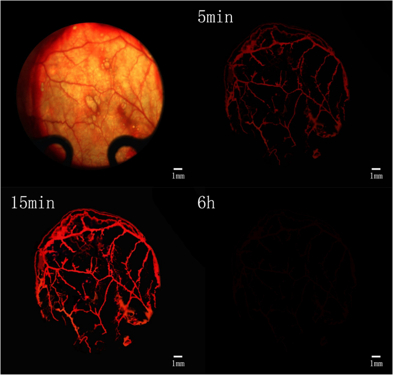
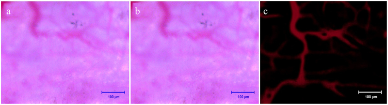
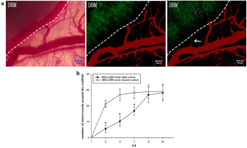
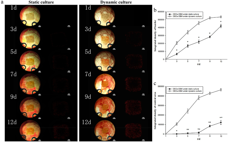
The current research on seed cells and scaffold materials of bone tissue engineering has achieved milestones. Nevertheless, necrosis of seed cells in center of bone scaffold is a bottleneck in tissue engineering. Therefore, this study aimed to investigate the inosculation mechanism of recipient microvasculature and prevascularized outgrowth endothelial progenitor cells (OECs)-demineralized bone matrix (DBM) complex. A dorsal skinfold window-chamber model with tail vein injection of Texas red-dextran was established to confirm the optimal observation time of microvessels. OECs-DBM complex under static and dynamic perfusion culture was implanted into the model to analyze vascularization. OECs-DBM complex was harvested on 12th day for HE staining and fluorescent imaging. The model was successfully constructed, and the most appropriate time to observe microvessels was 15 min after injection. The ingrowth of recipient microvessels arcoss the border of OECs-DBM complex increased with time in both groups, and more microvessels across the border were observed in dynamic perfusion group on 3rd, 5th, 7th day. Fluorescent integrated density of border in dynamic perfusion group was higher at all-time points, and the difference was more significant in central area. Fluorescent imaging of OECs-DBM complex exhibited that no enhanced green fluorescent protein-positive cells were found beyond the verge of DBM scaffold in both groups. prevascularization by dynamic perfusion culture can increase and accelerate the blood perfusion of OECs-DBM complex obtained from recipient microvasculature by internal inosculation. Accordingly, this approach may markedly contribute to the future success of tissue engineering applications in clinical practice.
目前,骨组织工程的种子细胞和支架材料的研究已经取得了里程碑式的进展。然而,种子细胞在骨支架中心的坏死是组织工程中的一个瓶颈。因此,本研究旨在探讨受体内皮祖细胞(EPCs)-脱钙骨基质(DBM)复合物与新生微血管吻合的机制。建立尾静脉注射 Texas Red-葡聚糖的背部皮肤囊窗室模型,以确认微血管的最佳观察时间。将 EPCs-DBM 复合物在静态和动态灌注培养条件下植入模型中,分析血管生成情况。在第 12 天收获 EPCs-DBM 复合物,进行 HE 染色和荧光成像。模型构建成功,注射后 15 min 是观察微血管的最佳时间。两组中,随着时间的推移,受体内皮细胞穿过 EPCs-DBM 复合物边界的血管向内生长增加,在动态灌注组的第 3、5、7 天观察到更多的血管穿过边界。动态灌注组边界的荧光积分密度在所有时间点均较高,且在中央区域差异更显著。EPCs-DBM 复合物的荧光成像显示,两组中均未在 DBM 支架的边缘以外发现增强型绿色荧光蛋白阳性细胞。动态灌注培养的预血管化可以增加和加速从受体内皮细胞获取的 EPCs-DBM 复合物的血液灌注。因此,这种方法可能对组织工程在临床实践中的未来成功应用有显著的贡献。