Hsu Huan-Yu, Chou Yu-Bai, Jheng Ying-Chun, Kao Zih-Kai, Huang Hsin-Yi, Chen Hung-Ruei, Hwang De-Kuang, Chen Shih-Jen, Chiou Shih-Hwa, Wu Yu-Te
Institute of Biophotonics, National Yang Ming Chiao Tung University, 155, Sec-2, Li Nong Street, Taipei 112304, Taiwan.
School of Medicine, National Yang Ming Chiao Tung University, Taipei 112304, Taiwan.
Biomedicines. 2022 May 29;10(6):1269. doi: 10.3390/biomedicines10061269.
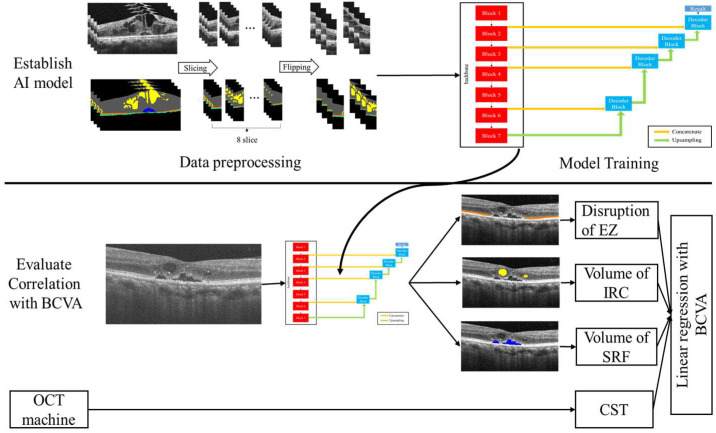
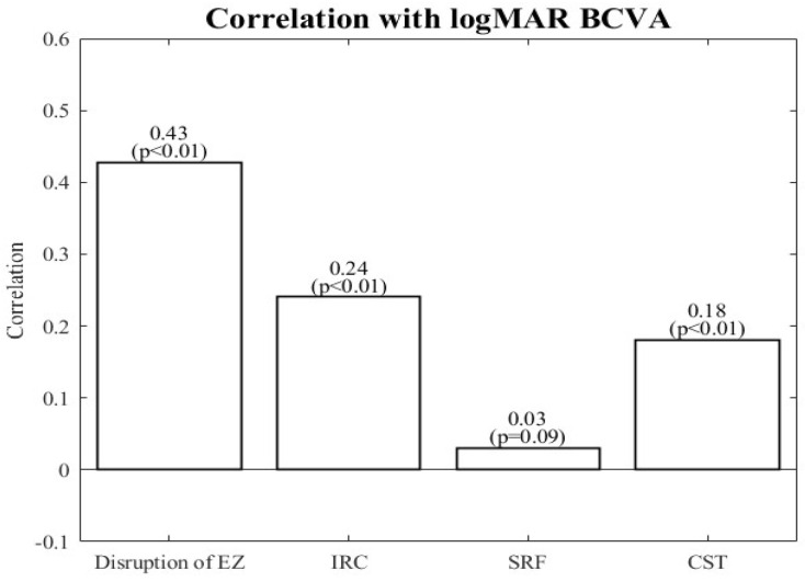
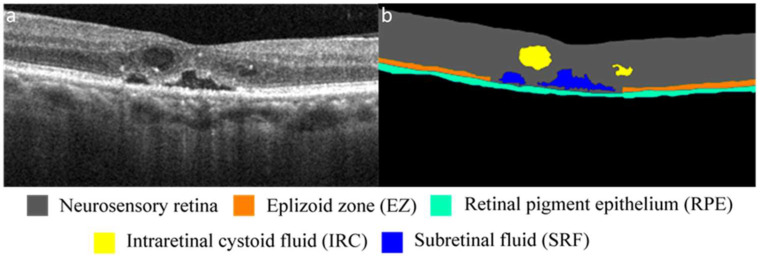
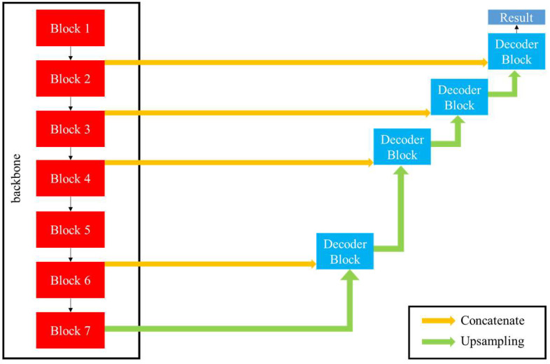
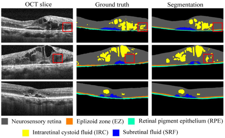
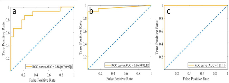
Diabetic macular edema (DME) is a highly common cause of vision loss in patients with diabetes. Optical coherence tomography (OCT) is crucial in classifying DME and tracking the results of DME treatment. The presence of intraretinal cystoid fluid (IRC) and subretinal fluid (SRF) and the disruption of the ellipsoid zone (EZ), which is part of the photoreceptor layer, are three crucial factors affecting the best corrected visual acuity (BCVA). However, the manual segmentation of retinal fluid and the EZ from retinal OCT images is laborious and time-consuming. Current methods focus only on the segmentation of retinal features, lacking a correlation with visual acuity. Therefore, we proposed a modified U-net, a deep learning algorithm, to segment these features from OCT images of patients with DME. We also correlated these features with visual acuity. The IRC, SRF, and EZ of the OCT retinal images were manually labeled and checked by doctors. We trained the modified U-net model on these labeled images. Our model achieved Sørensen-Dice coefficients of 0.80 and 0.89 for IRC and SRF, respectively. The area under the receiver operating characteristic curve (ROC) for EZ disruption was 0.88. Linear regression indicated that EZ disruption was the factor most strongly correlated with BCVA. This finding agrees with that of previous studies on OCT images. Thus, we demonstrate that our segmentation network can be feasibly applied to OCT image segmentation and assist physicians in assessing the severity of the disease.
糖尿病性黄斑水肿(DME)是糖尿病患者视力丧失的常见原因。光学相干断层扫描(OCT)对于DME的分类和追踪DME治疗结果至关重要。视网膜内囊样液(IRC)和视网膜下液(SRF)的存在以及作为光感受器层一部分的椭圆体带(EZ)的破坏是影响最佳矫正视力(BCVA)的三个关键因素。然而,从视网膜OCT图像中手动分割视网膜液和EZ既费力又耗时。当前方法仅专注于视网膜特征的分割,缺乏与视力的相关性。因此,我们提出了一种改进的U-net深度学习算法,用于从DME患者的OCT图像中分割这些特征。我们还将这些特征与视力相关联。OCT视网膜图像的IRC、SRF和EZ由医生手动标记并检查。我们在这些标记图像上训练改进的U-net模型。我们的模型对IRC和SRF的 Sørensen-Dice系数分别达到0.80和0.89。EZ破坏的受试者操作特征曲线(ROC)下面积为0.88。线性回归表明,EZ破坏是与BCVA相关性最强的因素。这一发现与先前关于OCT图像的研究一致。因此,我们证明我们的分割网络可以切实应用于OCT图像分割,并协助医生评估疾病的严重程度。