• 文献检索
  • 文档翻译
  • 深度研究
  • 学术资讯
  • Suppr Zotero 插件Zotero 插件
  • 邀请有礼
  • 套餐&价格
  • 历史记录
应用&插件
Suppr Zotero 插件Zotero 插件浏览器插件Mac 客户端Windows 客户端微信小程序
定价
高级版会员购买积分包购买API积分包
服务
文献检索文档翻译深度研究API 文档MCP 服务
关于我们
关于 Suppr公司介绍联系我们用户协议隐私条款
关注我们

Suppr 超能文献

核心技术专利:CN118964589B侵权必究
粤ICP备2023148730 号-1Suppr @ 2026

文献检索

告别复杂PubMed语法,用中文像聊天一样搜索,搜遍4000万医学文献。AI智能推荐,让科研检索更轻松。

立即免费搜索

文件翻译

保留排版,准确专业,支持PDF/Word/PPT等文件格式,支持 12+语言互译。

免费翻译文档

深度研究

AI帮你快速写综述,25分钟生成高质量综述,智能提取关键信息,辅助科研写作。

立即免费体验

健康年轻正常人眼底脉络膜毛细血管光相干断层扫描血管成像的计算机纹理分析。

Computerized Texture Analysis of Optical Coherence Tomography Angiography of Choriocapillaris in Normal Eyes of Young and Healthy Subjects.

机构信息

J. Terry Ernest Ocular Imaging Center, Department of Ophthalmology and Visual Science, The University of Chicago, Chicago, IL 60637, USA.

Department of Ophthalmology and Visual Science, University of Texas Health Sciences, Houston, TX 77030, USA.

出版信息

Cells. 2022 Jun 15;11(12):1934. doi: 10.3390/cells11121934.

DOI:10.3390/cells11121934
PMID:35741063
原文链接:https://pmc.ncbi.nlm.nih.gov/articles/PMC9221889/
Abstract

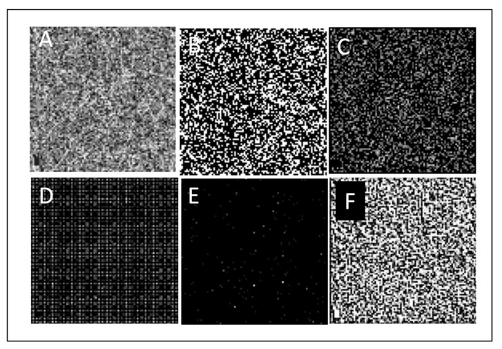
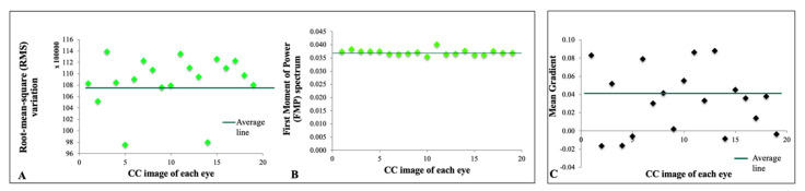
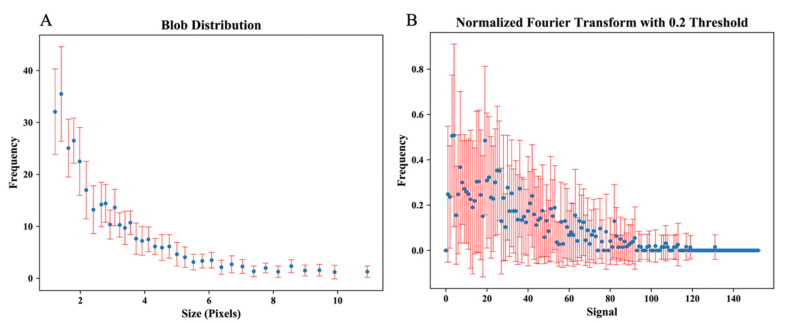
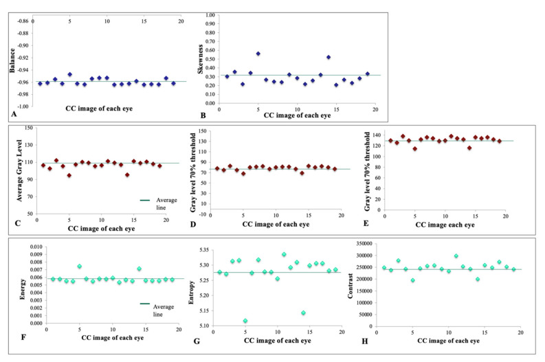
Computerized texture analysis uses higher-order mathematics to identify patterns beyond what the naked eye can recognize. We tested its feasibility in optical coherence tomography angiography imaging of choriocapillaris. Our objective was to determine sets of parameters that provide coherent and consistent output when applied to a homogeneous, healthy group of patients. This observational cross-sectional study involved 19 eyes of 10 young and healthy Caucasian subjects. En-face macular optical coherence tomography angiography of superficial choriocapillaris was obtained by the RTVue-XR Avanti system. Various algorithms were used to extract texture features. The mean and standard deviation were used to assess the distribution and dispersion of data points in each metric among eyes, which included: average gray level, gray level yielding 70% threshold and 30% threshold, balance, skewness, energy, entropy, contrast, edge mean gradient, root-mean-square variation, and first moment of power spectrum, which was compared between images, showing a highly concordant homology between all eyes of participants. We conclude that computerized texture analysis for en-face optical coherence tomography angiography images of choriocapillaris is feasible and provides values that are coherent and tightly distributed around the mean in a homogenous, healthy group of patients. Homology of blob size among subjects may represent a "repeat pattern" in signal density and thus a perfusion in the superficial choriocapillaris of healthy young individuals of the same ethnic background.

摘要

计算机纹理分析使用高阶数学来识别肉眼无法识别的模式。我们测试了它在脉络膜毛细血管光相干断层扫描血管造影成像中的可行性。我们的目的是确定在应用于同质、健康的患者群体时提供一致和一致输出的参数集。本观察性横断面研究涉及 10 名年轻健康白种人受试者的 19 只眼。通过 RTVue-XR Avanti 系统获得浅层脉络膜毛细血管的黄斑面光学相干断层扫描血管造影。使用各种算法提取纹理特征。平均值和标准差用于评估每个指标在眼睛之间的数据点的分布和离散程度,包括:平均灰度、产生 70%阈值和 30%阈值的灰度、平衡、偏度、能量、熵、对比度、边缘平均梯度、均方根变化和功率谱的第一矩,比较图像之间的差异,显示所有参与者的所有眼睛之间具有高度一致的同源性。我们得出结论,用于脉络膜毛细血管的面光学相干断层扫描血管造影图像的计算机纹理分析是可行的,并提供了在同质、健康的患者群体中一致且紧密分布在平均值周围的值。受试者中斑点大小的同源性可能代表信号密度中的“重复模式”,因此代表相同种族背景的健康年轻个体的浅层脉络膜毛细血管的灌注。

https://cdn.ncbi.nlm.nih.gov/pmc/blobs/6a0a/9221889/c3e52f896c51/cells-11-01934-g004.jpg
https://cdn.ncbi.nlm.nih.gov/pmc/blobs/6a0a/9221889/45ab4dd4f00d/cells-11-01934-g001.jpg
https://cdn.ncbi.nlm.nih.gov/pmc/blobs/6a0a/9221889/1bdddc161324/cells-11-01934-g002.jpg
https://cdn.ncbi.nlm.nih.gov/pmc/blobs/6a0a/9221889/8d5550ed6f4e/cells-11-01934-g003.jpg
https://cdn.ncbi.nlm.nih.gov/pmc/blobs/6a0a/9221889/c3e52f896c51/cells-11-01934-g004.jpg

相似文献

1
Computerized Texture Analysis of Optical Coherence Tomography Angiography of Choriocapillaris in Normal Eyes of Young and Healthy Subjects.健康年轻正常人眼底脉络膜毛细血管光相干断层扫描血管成像的计算机纹理分析。
Cells. 2022 Jun 15;11(12):1934. doi: 10.3390/cells11121934.
2
Choriocapillaris Imaging Using Multiple En Face Optical Coherence Tomography Angiography Image Averaging.使用多次表面光学相干断层扫描血管造影图像平均法进行脉络膜毛细血管成像
JAMA Ophthalmol. 2017 Nov 1;135(11):1197-1204. doi: 10.1001/jamaophthalmol.2017.3904.
3
Choriocapillaris flow deficit in a pachychoroid spectrum disease using en face optical coherence tomography angiography averaging.利用平均化脉络膜毛细血管血流亏损在厚脉络膜谱疾病中的应用。
PLoS One. 2022 Sep 12;17(9):e0271747. doi: 10.1371/journal.pone.0271747. eCollection 2022.
4
Concentric Choriocapillaris Flow Deficits in Retinitis Pigmentosa Detected Using Wide-Angle Swept-Source Optical Coherence Tomography Angiography.广角扫频源光相干断层血管成像检测到的视网膜色素变性的脉络膜毛细血管同心性血流缺损。
Invest Ophthalmol Vis Sci. 2019 Mar 1;60(4):1044-1049. doi: 10.1167/iovs.18-26176.
5
Choriocapillaris flow deficit in Bietti crystalline dystrophy detected using optical coherence tomography angiography.应用光相干断层扫描血管造影术检测先天性静止性夜盲症的脉络膜毛细血管血流不足。
Br J Ophthalmol. 2018 Sep;102(9):1208-1212. doi: 10.1136/bjophthalmol-2017-311313. Epub 2017 Dec 2.
6
Inter-relationship between retinal and choroidal vasculatures using optical coherence tomography angiography in normal eyes.正常眼视网膜与脉络膜血管系统之间的相互关系:光学相干断层扫描血管造影研究
Eur J Ophthalmol. 2020 Jan;30(1):48-57. doi: 10.1177/1120672118816225. Epub 2018 Dec 5.
7
Enhancement of Choriocapillaris Visualization in En Face Swept-Source Optical Coherence Tomography Angiography Images.增强脉络膜毛细血管在共面扫频源光相干断层扫描血管造影图像中的可视化。
Med Sci Monit. 2018 Jan 30;24:596-601. doi: 10.12659/msm.905534.
8
In Vivo Mapping of the Choriocapillaris in Healthy Eyes: A Widefield Swept-Source OCT Angiography Study.健康眼睛脉络膜毛细血管的体内成像:一项广角扫频源光学相干断层扫描血管造影研究。
Ophthalmol Retina. 2019 Nov;3(11):979-984. doi: 10.1016/j.oret.2019.05.026. Epub 2019 May 31.
9
Interaction Between the Distribution of Diabetic Retinopathy Lesions and the Association of Optical Coherence Tomography Angiography Scans With Diabetic Retinopathy Severity.糖尿病视网膜病变病变分布与光学相干断层扫描血管造影扫描与糖尿病视网膜病变严重程度的相关性分析。
JAMA Ophthalmol. 2020 Dec 1;138(12):1291-1297. doi: 10.1001/jamaophthalmol.2020.4516.
10
Evaluation of the choroidal features in pachychoroid spectrum diseases by optical coherence tomography and optical coherence tomography angiography.利用光学相干断层扫描和光学相干断层扫描血管成像评估肥厚脉络膜谱疾病的脉络膜特征。
Eur J Ophthalmol. 2021 Jan;31(1):184-193. doi: 10.1177/1120672119887095. Epub 2019 Nov 4.

引用本文的文献

1
Assessment of Choriocapillaris in Healthy and Diseased Eyes Using 3-Dimensional Topographic Maps Based on OCT Angiography.基于光学相干断层扫描血管造影的三维地形图对健康和患病眼睛脉络膜毛细血管的评估
Ophthalmol Sci. 2025 May 17;5(6):100828. doi: 10.1016/j.xops.2025.100828. eCollection 2025 Nov-Dec.
2
Radiomics-Based OCT Analysis of Choroid Reveals Biomarkers of Central Serous Chorioretinopathy.基于影像组学的脉络膜光学相干断层扫描分析揭示了中心性浆液性脉络膜视网膜病变的生物标志物。
Transl Vis Sci Technol. 2025 Apr 1;14(4):23. doi: 10.1167/tvst.14.4.23.
3
Radiomics-Based Prediction of Anti-VEGF Treatment Response in Neovascular Age-Related Macular Degeneration With Pigment Epithelial Detachment.

本文引用的文献

1
1.7-Micron Optical Coherence Tomography Angiography for Characterization of Skin Lesions-A Feasibility Study.1.7 微米光学相干断层扫描血管造影术用于皮肤病变的特征分析:一项可行性研究。
IEEE Trans Med Imaging. 2021 Sep;40(9):2507-2512. doi: 10.1109/TMI.2021.3081066. Epub 2021 Aug 31.
2
Identification and Characterization of Epivascular Glia Using En Face Optical Coherence Tomography.使用共聚焦光学相干断层扫描技术鉴定和描述血管周细胞。
Am J Ophthalmol. 2021 Sep;229:108-119. doi: 10.1016/j.ajo.2021.03.014. Epub 2021 Mar 24.
3
Evaluation of the different thresholding strategies for quantifying choriocapillaris using optical coherence tomography angiography.
基于影像组学对伴有色素上皮脱离的新生血管性年龄相关性黄斑变性抗VEGF治疗反应的预测
Transl Vis Sci Technol. 2023 Oct 3;12(10):3. doi: 10.1167/tvst.12.10.3.
4
Racial differences in quantitative optical coherence tomography angiography findings between older non-diabetics with co-morbidities.老年人合并症中非糖尿病患者定量光学相干断层扫描血管造影结果的种族差异。
PLoS One. 2023 May 5;18(5):e0285360. doi: 10.1371/journal.pone.0285360. eCollection 2023.
使用光学相干断层扫描血管造影术评估用于量化脉络膜毛细血管的不同阈值策略。
Quant Imaging Med Surg. 2020 Oct;10(10):1994-2005. doi: 10.21037/qims-20-340.
4
Multiple scan averaging to yield accurate quantitative analysis of optical coherence tomography angiograms.多次扫描平均以获得光相干断层扫描血管造影的准确定量分析。
Sci Rep. 2020 Apr 10;10(1):6194. doi: 10.1038/s41598-020-62956-2.
5
Topographic analysis of macular choriocapillaris flow deficits in diabetic retinopathy using swept-source optical coherence tomography angiography.使用扫频源光学相干断层扫描血管造影术对糖尿病视网膜病变中黄斑脉络膜毛细血管血流缺损进行的地形图分析。
Int J Retina Vitreous. 2020 Mar 19;6:6. doi: 10.1186/s40942-020-00209-0. eCollection 2020.
6
Quantification of Choriocapillaris with Phansalkar Local Thresholding: Pitfalls to Avoid.用 Phansalkar 局部阈值法对脉络膜毛细血管进行定量分析:应避免的陷阱。
Am J Ophthalmol. 2020 May;213:161-176. doi: 10.1016/j.ajo.2020.02.003. Epub 2020 Feb 12.
7
Correlations Between Different Choriocapillaris Flow Deficit Parameters in Normal Eyes Using Swept Source OCT Angiography.应用扫频源 OCT 血管造影术评估正常眼中不同脉络膜血流缺损参数之间的相关性。
Am J Ophthalmol. 2020 Jan;209:18-26. doi: 10.1016/j.ajo.2019.09.017. Epub 2019 Sep 25.
8
Quantification of Choriocapillaris with Optical Coherence Tomography Angiography: A Comparison Study.脉络膜毛细血管的光学相干断层扫描血管造影定量分析:一项对比研究。
Am J Ophthalmol. 2019 Dec;208:111-123. doi: 10.1016/j.ajo.2019.07.003. Epub 2019 Jul 16.
9
Correlations between Choriocapillaris Flow Deficits around Geographic Atrophy and Enlargement Rates Based on Swept-Source OCT Imaging.基于扫频源光学相干断层扫描成像的地图样萎缩周围脉络膜毛细血管血流缺损与扩大率之间的相关性
Ophthalmol Retina. 2019 Jun;3(6):478-488. doi: 10.1016/j.oret.2019.01.024. Epub 2019 Feb 6.
10
Quantitative Assessment of Choriocapillaris Flow Deficits in Eyes with Advanced Age-Related Macular Degeneration Versus Healthy Eyes.定量评估晚期年龄相关性黄斑变性眼与健康眼的脉络膜毛细血管血流缺损。
Am J Ophthalmol. 2019 Sep;205:132-139. doi: 10.1016/j.ajo.2019.04.037. Epub 2019 May 10.