Sándor-Bajusz Kinga A, Sadi Asaad, Varga Eszter, Csábi Györgyi, Antonoglou Georgios N, Lohner Szimonetta
Department of Pediatrics, University of Pécs, Pécs, Hungary.
Doctoral School of Clinical Neurosciences, University of Pécs, Pécs, Hungary.
Front Neuroanat. 2022 Jun 10;16:863900. doi: 10.3389/fnana.2022.863900. eCollection 2022.
Neuroimaging of individuals with non-syndromic oral clefts have revealed subtle brain structural differences compared to matched controls. Previous studies strongly suggest a unified primary dysfunction of normal brain and face development which could explain these neuroanatomical differences and the neuropsychiatric issues frequently observed in these individuals. Currently there are no studies that have assessed the overall empirical evidence of the association between oral clefts and brain structure. Our aim was to summarize the available evidence on potential brain structural differences in individuals with non-syndromic oral clefts and their matched controls.
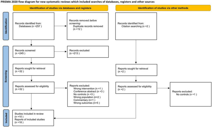
MEDLINE, Scopus, Cochrane Central Register of Controlled Trials, Web of Science and Embase were systematically searched in September 2020 for case-control studies that reported structural brain MRI in individuals with non-syndromic oral clefts and healthy controls. Studies of syndromic oral clefts were excluded. Two review authors independently screened studies for eligibility, extracted data and assessed risk of bias with the Newcastle-Ottawa Scale. Random effects meta-analyses of mean differences (MDs) and their 95% confidence intervals (95% CI) were performed in order to compare global and regional brain MRI volumes.
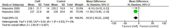
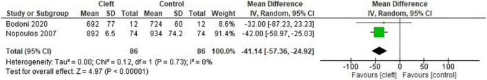
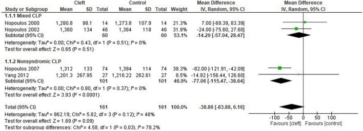
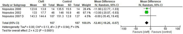
Ten studies from 18 records were included in the review. A total of 741 participants were analyzed. A moderate to high risk of bias was determined for the included studies. The cerebellum (MD: -12.46 cm, 95% CI: -18.26, -6.67, = 3 studies, 354 participants), occipital lobes (MD: -7.39, 95% CI: -12.80, -1.99, = 2 studies, 120 participants), temporal lobes (MD: -10.53 cm, 95% CI: -18.23, -2.82, = 2 studies, 120 participants) and total gray matter (MD: -41.14 cm; 95% CI: -57.36 to -24.92, = 2 studies, 172 participants) were significantly smaller in the cleft group compared to controls.
There may be structural brain differences between individuals with non-syndromic oral clefts and controls based on the available evidence. Improvement in study design, size, methodology and participant selection could allow a more thorough analysis and decrease study heterogeneity.
与匹配的对照组相比,非综合征性口腔颌面部裂隙患者的神经影像学检查显示出细微的脑结构差异。先前的研究强烈表明,正常脑和面部发育存在统一的原发性功能障碍,这可以解释这些神经解剖学差异以及这些患者中经常观察到的神经精神问题。目前尚无研究评估口腔颌面部裂隙与脑结构之间关联的总体实证证据。我们的目的是总结关于非综合征性口腔颌面部裂隙患者及其匹配对照组潜在脑结构差异的现有证据。
2020年9月,我们系统检索了MEDLINE、Scopus、Cochrane对照试验中央注册库、科学网和Embase,以查找报告非综合征性口腔颌面部裂隙患者和健康对照者脑结构MRI的病例对照研究。排除综合征性口腔颌面部裂隙的研究。两位综述作者独立筛选研究的纳入资格,提取数据并使用纽卡斯尔-渥太华量表评估偏倚风险。为了比较全脑和局部脑MRI体积,我们进行了随机效应荟萃分析,计算平均差异(MD)及其95%置信区间(95%CI)。
本综述纳入了18条记录中的10项研究。共分析了741名参与者。纳入的研究存在中度至高度偏倚风险。与对照组相比,裂隙组的小脑(MD:-12.46cm,95%CI:-18.26,-6.67;n = 3项研究,354名参与者)、枕叶(MD:-7.39,95%CI:-12.80,-1.99;n = 2项研究,120名参与者)、颞叶(MD:-10.53cm,95%CI:-18.23,-2.82;n = 2项研究,120名参与者)和总灰质(MD:-41.14cm;95%CI:-57.36至-24.92;n = 2项研究,172名参与者)明显较小。
根据现有证据,非综合征性口腔颌面部裂隙患者与对照组之间可能存在脑结构差异。改进研究设计、规模、方法和参与者选择可以进行更全面的分析并减少研究异质性。