Lexington Veterans' Affairs Healthcare System, Lexington, KY, USA.
Department of Physiology, University of Kentucky, Lexington, USA.
Acta Neuropathol Commun. 2022 Jun 27;10(1):93. doi: 10.1186/s40478-022-01395-8.
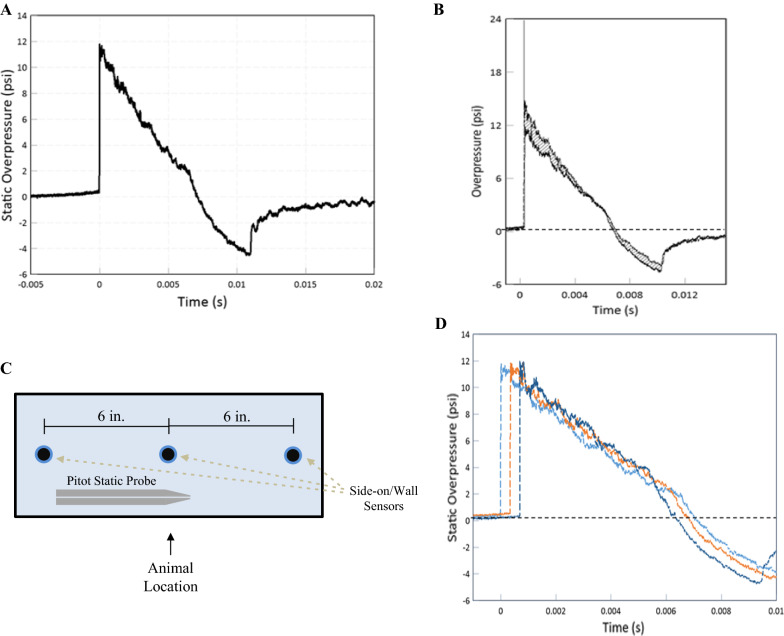
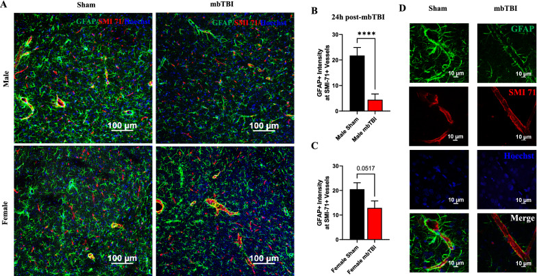
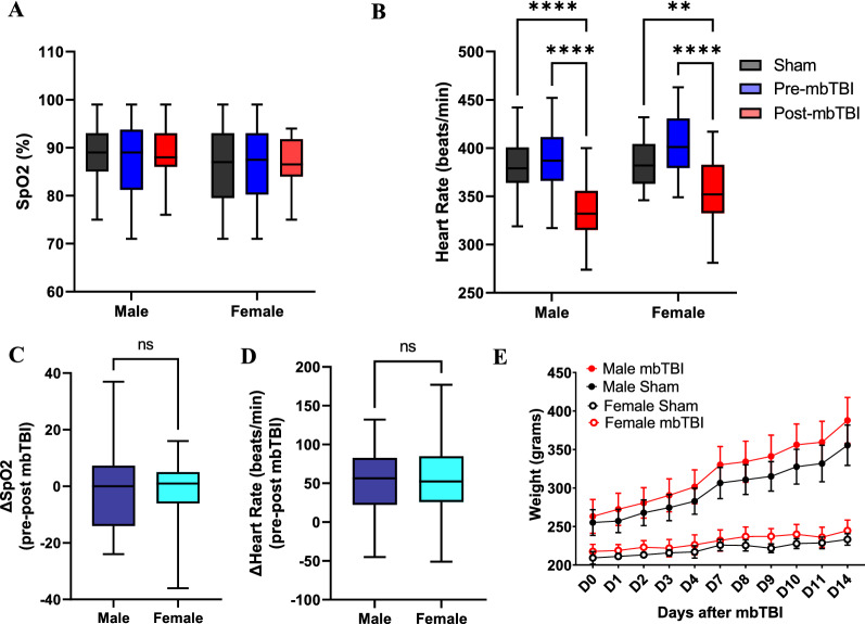
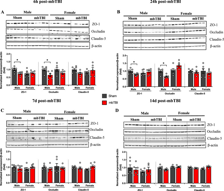
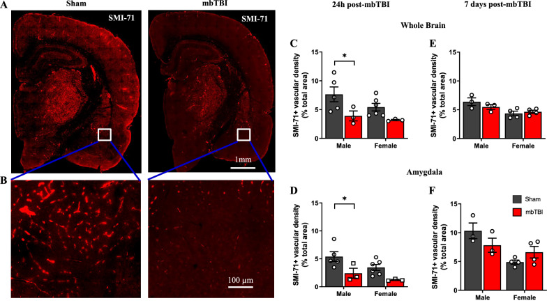
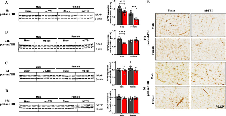
Low-level blast exposure can result in neurological impairment for military personnel. Currently, there is a lack of experimental data using sex as a biological variable in neurovascular outcomes following blast exposure. To model mild blast traumatic brain injury (mbTBI), male and female rats were exposed to a single 11 psi static peak overpressure blast wave using the McMillan blast device and cohorts were then euthanized at 6 h, 24 h, 7 d and 14 d post-blast followed by isolation of the amygdala. After mbTBI, animals experience immediate bradycardia, although no changes in oxygen saturation levels or weight loss are observed. Male mbTBI animals displayed significantly higher levels of anxiety-like behavior (open field and elevated plus maze) compared to male sham groups; however, there was no anxiety phenotype in female mbTBI animals. Blast-induced neurovascular damage was explored by measuring expression of tight junction (TJ) proteins (zonula occludens-1 (ZO-1), occludin and claudin-5), glial fibrillary acidic protein (GFAP) and astrocyte end-feet coverage around the blood-brain barrier (BBB). Western blot analysis demonstrates that TJ protein levels were significantly decreased at 6 h and 24 h post-mbTBI in male rats, but not in female rats, compared to sham. Female animals have decreased GFAP at 6 h post-mbTBI while male animals display decreased GFAP expression at 24 h post-mbTBI. By 7 d post-mbTBI, there were no significant differences in TJ or GFAP levels between groups in either sex. At 24 h post-mbTBI, vascular integrity and astrocytic end-feet coverage around the BBB was significantly decreased in males following mbTBI. These results demonstrate that loss of GFAP expression may be due to astrocytic damage at the BBB. Our findings also demonstrate sex differences in acute vascular and behavioral outcomes after single mbTBI. Female animals display a lack of BBB pathology after mbTBI corresponding to improved acute neuropsychological outcomes as compared to male animals.
低水平爆炸暴露可导致军事人员的神经损伤。目前,在爆炸暴露后神经血管结果中,缺乏使用性别作为生物学变量的实验数据。为了模拟轻度爆炸性脑损伤(mbTBI),雄性和雌性大鼠使用 McMillan 爆炸装置暴露于单个 11psi 静态峰值超压爆炸波中,然后在爆炸后 6h、24h、7d 和 14d 处死队列,随后分离杏仁核。在 mbTBI 后,动物立即出现心动过缓,尽管未观察到氧饱和度水平或体重减轻的变化。与雄性假手术组相比,雄性 mbTBI 动物表现出明显更高水平的焦虑样行为(旷场和高架十字迷宫);然而,雌性 mbTBI 动物没有焦虑表型。通过测量紧密连接(TJ)蛋白(闭合蛋白-1(ZO-1)、occludin 和 claudin-5)、胶质纤维酸性蛋白(GFAP)和血脑屏障(BBB)周围星形胶质细胞终足覆盖的表达来探索爆炸诱导的神经血管损伤。Western blot 分析表明,与假手术相比,雄性大鼠 mbTBI 后 6h 和 24h TJ 蛋白水平显著降低,但雌性大鼠则没有。雌性动物在 mbTBI 后 6h 时 GFAP 减少,而雄性动物在 mbTBI 后 24h 时 GFAP 表达减少。在 mbTBI 后 7d,两组在任性别中 TJ 或 GFAP 水平均无显著差异。在 mbTBI 后 24h,雄性动物的血管完整性和 BBB 周围星形胶质细胞终足覆盖明显减少。这些结果表明,GFAP 表达的丧失可能是由于 BBB 处的星形胶质细胞损伤。我们的研究结果还表明,单次 mbTBI 后急性血管和行为结果存在性别差异。与雄性动物相比,mbTBI 后雌性动物的 BBB 病理缺失,对应于急性神经心理学结局的改善。