• 文献检索
  • 文档翻译
  • 深度研究
  • 学术资讯
  • Suppr Zotero 插件Zotero 插件
  • 邀请有礼
  • 套餐&价格
  • 历史记录
应用&插件
Suppr Zotero 插件Zotero 插件浏览器插件Mac 客户端Windows 客户端微信小程序
定价
高级版会员购买积分包购买API积分包
服务
文献检索文档翻译深度研究API 文档MCP 服务
关于我们
关于 Suppr公司介绍联系我们用户协议隐私条款
关注我们

Suppr 超能文献

核心技术专利:CN118964589B侵权必究
粤ICP备2023148730 号-1Suppr @ 2026

文献检索

告别复杂PubMed语法,用中文像聊天一样搜索,搜遍4000万医学文献。AI智能推荐,让科研检索更轻松。

立即免费搜索

文件翻译

保留排版,准确专业,支持PDF/Word/PPT等文件格式,支持 12+语言互译。

免费翻译文档

深度研究

AI帮你快速写综述,25分钟生成高质量综述,智能提取关键信息,辅助科研写作。

立即免费体验

住院 SARS-CoV-2 感染患者的胸部 CT 表现:Delta 变异株与奥密克戎变异株。

Chest CT Findings in Hospitalized Patients with SARS-CoV-2: Delta versus Omicron Variants.

机构信息

From the Department of Radiology, Seoul National University Hospital, Seoul National University College of Medicine, Seoul, Korea (S.H.Y., J.H.L.); and Department of Internal Medicine, Inje University Sanggye Paik Hospital, Inje University College of Medicine, Seoul 01757, Korea (B.N.K.).

出版信息

Radiology. 2023 Jan;306(1):252-260. doi: 10.1148/radiol.220676. Epub 2022 Jun 28.

DOI:10.1148/radiol.220676
PMID:35762887
原文链接:https://pmc.ncbi.nlm.nih.gov/articles/PMC9272824/
Abstract

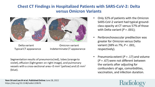
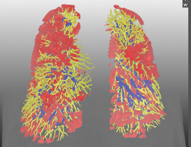
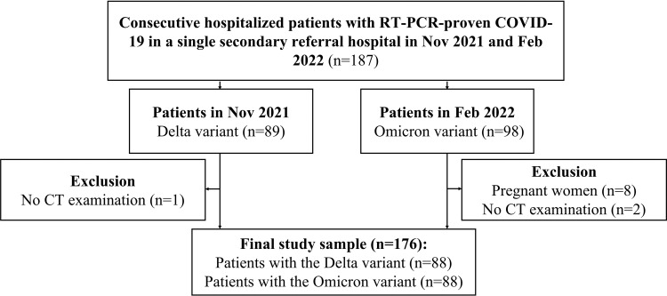
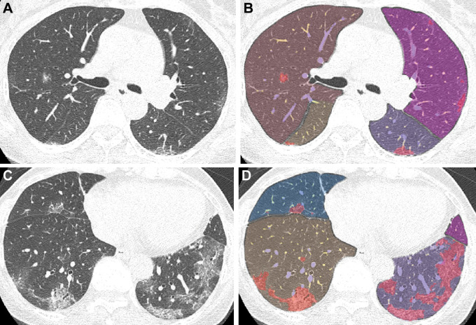
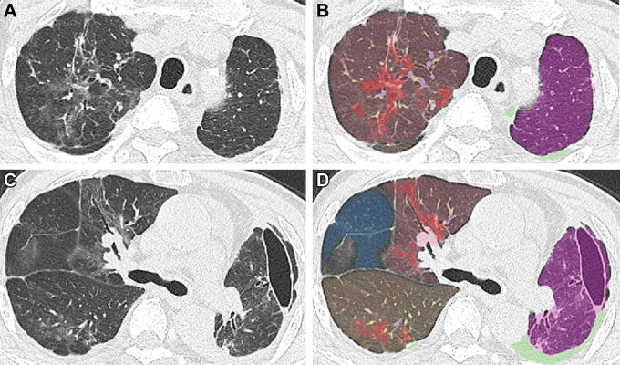
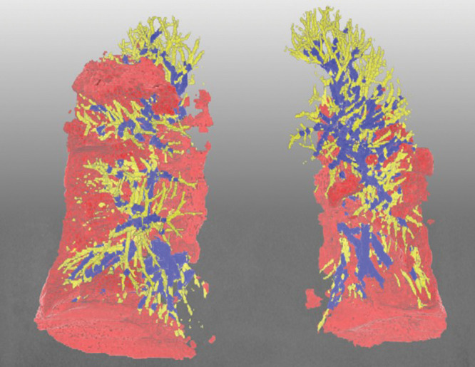
Background CT manifestations of SARS-CoV-2 may differ among variants. Purpose To compare the chest CT findings of SARS-CoV-2 between the Delta and Omicron variants. Materials and Methods This retrospective study collected consecutive baseline chest CT images of hospitalized patients with SARS-CoV-2 from a secondary referral hospital when the Delta and Omicron variants were predominant. Two radiologists categorized CT images according to the RSNA classification system for COVID-19 and visually graded pneumonia extent. Pneumonia, pleural effusion, and intrapulmonary vessels were segmented and quantified on CT images using a priori-developed neural networks, followed by reader confirmation. Multivariable logistic and linear regression analyses were performed to examine the associations between the variants and CT category, distribution, severity, and peripheral vascularity. Results In total, 88 patients with the Delta variant (mean age, 67 years ± 15 [SD]; 46 men) and 88 patients with the Omicron variant (mean age, 62 years ± 19; 51 men) were included. Omicron was associated with less frequent, typical peripheral bilateral ground-glass opacity (32% [28 of 88] vs 57% [50 of 88], = .001), more frequent peribronchovascular predilection (38% [25 of 66] vs 7% [five of 71], < .001), lower visual pneumonia extent (5.4 ± 6.0 vs 7.7 ± 6.6, = .02), similar pneumonia volume (5% ± 1 vs 7% ± 11, = .14), and a higher proportion of vessels with a cross-sectional area smaller than 5 mm relative to the total pulmonary blood volume (BV5%; 48% ± 11 vs 44% ± 8; = .004). In adjusted analyses, Omicron was associated with a nontypical appearance (odds ratio, 0.34; = .006), peribronchovascular predilection (odds ratio, 9.2; < .001), and higher BV5% (β = 3.8; = .01) but similar visual pneumonia extent ( = .17) and pneumonia volume ( = .67) relative to the Delta variant. Conclusion At chest CT, the Omicron SARS-COV-2 variant showed nontypical peribronchovascular pneumonia and less pulmonary vascular involvement than did the Delta variant in hospitalized patients with similar disease severity. © RSNA, 2022

摘要

背景

SARS-CoV-2 的 CT 表现可能因变体而异。目的:比较德尔塔和奥密克戎变体的 SARS-CoV-2 之间的胸部 CT 发现。材料与方法:本回顾性研究收集了一家二级转诊医院住院的 SARS-CoV-2 患者的连续基线胸部 CT 图像,当时德尔塔和奥密克戎变体占主导地位。两位放射科医生根据 RSNA 分类系统对 CT 图像进行分类,并对肺炎程度进行视觉分级。使用预先开发的神经网络对 CT 图像上的肺炎、胸腔积液和肺内血管进行分割和量化,然后由读者确认。进行多变量逻辑和线性回归分析,以检查变体与 CT 类别、分布、严重程度和周围血管性之间的关联。结果:共纳入 88 例德尔塔变体患者(平均年龄,67 岁±15[标准差];46 名男性)和 88 例奥密克戎变体患者(平均年龄,62 岁±19;51 名男性)。奥密克戎与更常见的非典型双侧外周性磨玻璃影(32%[28/88]与 57%[50/88],.001)、更常见的支气管周围血管偏好(38%[25/66]与 7%[5/71], <.001)、较低的视觉肺炎程度(5.4±6.0 与 7.7±6.6,.02)、相似的肺炎体积(5%±1 与 7%±11, =.14)和更大比例的截面积小于总肺血容积(BV5%)的血管(48%±11 与 44%±8, =.004)有关。在调整分析中,奥密克戎与非典型表现(比值比,0.34;.006)、支气管周围血管偏好(比值比,9.2; <.001)和更高的 BV5%(β=3.8;.01)相关,但与德尔塔变体相比,视觉肺炎程度( =.17)和肺炎体积( =.67)相似。结论:在胸部 CT 上,与德尔塔变体相比,住院患者中奥密克戎 SARS-COV-2 变体表现为非典型性支气管周围性肺炎,肺血管受累程度较低,且疾病严重程度相似。

https://cdn.ncbi.nlm.nih.gov/pmc/blobs/d5fa/9792714/0ff6d63f84cb/radiol.220676.fig5.jpg
https://cdn.ncbi.nlm.nih.gov/pmc/blobs/d5fa/9792714/5aa03a942c2b/radiol.220676.VA.jpg
https://cdn.ncbi.nlm.nih.gov/pmc/blobs/d5fa/9792714/8cdbbbbf8b71/radiol.220676.fig1.jpg
https://cdn.ncbi.nlm.nih.gov/pmc/blobs/d5fa/9792714/f60ae7024499/radiol.220676.fig2.jpg
https://cdn.ncbi.nlm.nih.gov/pmc/blobs/d5fa/9792714/226d2a05e26a/radiol.220676.fig3.jpg
https://cdn.ncbi.nlm.nih.gov/pmc/blobs/d5fa/9792714/51dfcf43bab0/radiol.220676.fig4.jpg
https://cdn.ncbi.nlm.nih.gov/pmc/blobs/d5fa/9792714/0ff6d63f84cb/radiol.220676.fig5.jpg

相似文献

1
Chest CT Findings in Hospitalized Patients with SARS-CoV-2: Delta versus Omicron Variants.住院 SARS-CoV-2 感染患者的胸部 CT 表现:Delta 变异株与奥密克戎变异株。
Radiology. 2023 Jan;306(1):252-260. doi: 10.1148/radiol.220676. Epub 2022 Jun 28.
2
Reduction in Chest CT Severity and Improved Hospital Outcomes in SARS-CoV-2 Omicron Compared with Delta Variant Infection.与德尔塔变异株感染相比,奥密克戎变异株感染导致胸部 CT 严重程度降低和住院结局改善。
Radiology. 2023 Jan;306(1):261-269. doi: 10.1148/radiol.220533. Epub 2022 Jun 21.
3
Impact of Vaccination and the Omicron Variant on COVID-19-related Chest CT Findings: A Multicenter Study.疫苗接种和奥密克戎变异株对 COVID-19 相关胸部 CT 表现的影响:一项多中心研究。
Radiology. 2023 May;307(3):e222730. doi: 10.1148/radiol.222730. Epub 2023 Mar 7.
4
SARS-CoV-2 Variants Infection in Relationship to Imaging-based Pneumonia and Clinical Outcomes.SARS-CoV-2 变体感染与基于影像学的肺炎及临床结局的关系。
Radiology. 2023 Mar;306(3):e221795. doi: 10.1148/radiol.221795. Epub 2022 Sep 27.
5
Comparison between original SARS-CoV-2 strain and omicron variant on thin-section chest CT imaging of COVID-19 pneumonia.比较新冠肺炎薄层层析 CT 影像中原始 SARS-CoV-2 株与奥密克戎变异株。
Radiologie (Heidelb). 2023 Nov;63(Suppl 2):55-63. doi: 10.1007/s00117-023-01147-2. Epub 2023 Jun 6.
6
Real life treatment experience and outcome of consecutively hospitalised patients with SARS-CoV-2 pneumonia by Omicron-1 vs Delta variants.奥密克戎-1 变异株与德尔塔变异株所致 SARS-CoV-2 肺炎患者连续住院的真实世界治疗经验和结局。
Infect Dis (Lond). 2023 Oct;55(10):706-715. doi: 10.1080/23744235.2023.2232445. Epub 2023 Jul 10.
7
Rate of True-Positive Findings of COVID-19 Typical Appearance at Chest CT per RSNA Consensus Guidelines in an Increasingly Vaccinated Population.在接种疫苗人数不断增加的情况下,根据 RSNA 共识指南,胸部 CT 显示 COVID-19 典型表现的真阳性发现率。
Radiology. 2023 Mar;306(3):e220680. doi: 10.1148/radiol.220680. Epub 2022 Sep 6.
8
Computed Tomographic Imaging Features of COVID-19 Pneumonia Caused by the Delta (B.1.617.2) and Omicron (B.1.1.529) Variant in a German Nested Cohort Pilot Study Group.德国嵌套队列试点研究组中德尔塔(B.1.617.2)和奥密克戎(B.1.1.529)变异株引起的新冠肺炎的计算机断层扫描成像特征。
Tomography. 2022 Sep 28;8(5):2435-2449. doi: 10.3390/tomography8050202.
9
Survey on chest CT findings in COVID-19 patients in Okinawa, Japan: differences between the delta and omicron variants.日本冲绳地区 COVID-19 患者的胸部 CT 表现调查:德尔塔和奥密克戎变异株的差异。
Sci Rep. 2023 Nov 21;13(1):20373. doi: 10.1038/s41598-023-47756-8.
10
Comparison of Clinical Outcomes and Imaging Features in Hospitalized Patients with SARS-CoV-2 Omicron Subvariants.奥密克戎变异株亚分支感染住院患者的临床结局和影像学特征比较。
Radiology. 2023 Jul;308(1):e230653. doi: 10.1148/radiol.230653.

引用本文的文献

1
Nanomaterial-Based Biosensors for the Detection of COVID-19.用于检测新型冠状病毒肺炎的纳米材料基生物传感器
Indian J Microbiol. 2025 Mar;65(1):120-136. doi: 10.1007/s12088-024-01336-0. Epub 2024 Jun 23.
2
Differences in chest imaging between Omicron and non-Omicron coronavirus disease 2019 (COVID-19) patients: a systematic review and meta-analysis.奥密克戎与非奥密克戎新型冠状病毒肺炎(COVID-19)患者胸部影像学差异:一项系统评价与荟萃分析
BMC Infect Dis. 2025 Apr 29;25(1):631. doi: 10.1186/s12879-025-11032-z.
3
Risk factors for SARS-CoV-2 pneumonia among renal transplant recipients in Omicron pandemic-a prospective cohort study.

本文引用的文献

1
Deep Learning-Based Automatic CT Quantification of Coronavirus Disease 2019 Pneumonia: An International Collaborative Study.基于深度学习的 2019 年冠状病毒病肺炎 CT 定量分析:一项国际合作研究。
J Comput Assist Tomogr. 2022;46(3):413-422. doi: 10.1097/RCT.0000000000001303. Epub 2022 Apr 8.
2
Estimates of SARS-CoV-2 Omicron Variant Severity in Ontario, Canada.加拿大安大略省估计 SARS-CoV-2 奥密克戎变异株的严重程度。
JAMA. 2022 Apr 5;327(13):1286-1288. doi: 10.1001/jama.2022.2274.
3
SARS-CoV-2 Omicron variant replication in human bronchus and lung ex vivo.
奥密克戎大流行期间肾移植受者感染新型冠状病毒肺炎的危险因素——一项前瞻性队列研究
Virol J. 2024 Dec 4;21(1):315. doi: 10.1186/s12985-024-02591-9.
4
Evaluating the Accuracy of Chest CT in Detecting COVID-19 Through Tracheobronchial Wall Thickness: Insights From Emergency Department Patients in Mid-2023.通过气管支气管壁厚度评估胸部CT检测2023年年中急诊科患者新型冠状病毒肺炎的准确性:见解
Cureus. 2024 Sep 11;16(9):e69161. doi: 10.7759/cureus.69161. eCollection 2024 Sep.
5
Vascular Pathogenesis in Acute and Long COVID: Current Insights and Therapeutic Outlook.急性和长期新冠病毒感染中的血管发病机制:当前见解与治疗前景
Semin Thromb Hemost. 2025 Apr;51(3):256-271. doi: 10.1055/s-0044-1790603. Epub 2024 Sep 30.
6
Physiological and Proteomic Analysis of Various Priming on Rice Seed under Chilling Stress.低温胁迫下不同引发处理对水稻种子的生理和蛋白质组学分析
Plants (Basel). 2024 Aug 30;13(17):2430. doi: 10.3390/plants13172430.
7
Omicron variant and pulmonary involvements: a chest imaging analysis in asymptomatic and mild COVID-19.奥密克戎变异株与肺部受累:无症状和轻症 COVID-19 的胸部影像学分析。
Front Public Health. 2024 Jul 4;12:1325474. doi: 10.3389/fpubh.2024.1325474. eCollection 2024.
8
Salivary biomarkers as pioneering indicators for diagnosis and severity stratification of pediatric long COVID.唾液生物标志物可作为儿科长新冠诊断和严重程度分层的开创性指标。
Front Cell Infect Microbiol. 2024 May 31;14:1396263. doi: 10.3389/fcimb.2024.1396263. eCollection 2024.
9
Comparison of Computed Tomography and Clinical Features Between Patients Infected with the SARS-CoV-2 Omicron Variant and the Original Strain.感染严重急性呼吸综合征冠状病毒2(SARS-CoV-2)奥密克戎变异株与原始毒株患者的计算机断层扫描及临床特征比较
Infect Drug Resist. 2024 Mar 5;17:807-818. doi: 10.2147/IDR.S448713. eCollection 2024.
10
The evolution of lung computed tomography findings in COVID-19 from 2020 to 2023: more signs of co-infection.2020年至2023年COVID-19患者肺部计算机断层扫描结果的演变:合并感染迹象增多。
ERJ Open Res. 2024 Feb 26;10(1). doi: 10.1183/23120541.00727-2023. eCollection 2024 Jan.
严重急性呼吸综合征冠状病毒2型奥密克戎变异株在人支气管和肺组织外植体中的复制
Nature. 2022 Mar;603(7902):715-720. doi: 10.1038/s41586-022-04479-6. Epub 2022 Feb 1.
4
Imaging and Clinical Features of COVID-19 Breakthrough Infections: A Multicenter Study.COVID-19 突破性感染的影像学和临床特征:一项多中心研究。
Radiology. 2022 Jun;303(3):682-692. doi: 10.1148/radiol.213072. Epub 2022 Feb 1.
5
Trends in Disease Severity and Health Care Utilization During the Early Omicron Variant Period Compared with Previous SARS-CoV-2 High Transmission Periods - United States, December 2020-January 2022.疾病严重程度和医疗保健利用趋势在奥密克戎变异株早期与之前 SARS-CoV-2 高传播期相比-美国,2020 年 12 月至 2022 年 1 月。
MMWR Morb Mortal Wkly Rep. 2022 Jan 28;71(4):146-152. doi: 10.15585/mmwr.mm7104e4.
6
Viral infection and transmission in a large, well-traced outbreak caused by the SARS-CoV-2 Delta variant.新冠病毒德尔塔变异株引发的大型溯源清晰的暴发中的病毒感染和传播。
Nat Commun. 2022 Jan 24;13(1):460. doi: 10.1038/s41467-022-28089-y.
7
Early assessment of the clinical severity of the SARS-CoV-2 omicron variant in South Africa: a data linkage study.南非对 SARS-CoV-2 奥密克戎变异株临床严重程度的早期评估:一项数据关联研究。
Lancet. 2022 Jan 29;399(10323):437-446. doi: 10.1016/S0140-6736(22)00017-4. Epub 2022 Jan 19.
8
Covid-19: We have good treatments for omicron, but questions remain, say doctors.医生表示,新冠疫情:我们对奥密克戎有有效的治疗方法,但问题仍然存在。
BMJ. 2022 Jan 11;376:o61. doi: 10.1136/bmj.o61.
9
Severity of Illness in Persons Infected With the SARS-CoV-2 Delta Variant vs Beta Variant in Qatar.在卡塔尔感染 SARS-CoV-2 的德尔塔变异株和贝塔变异株患者的疾病严重程度。
JAMA Intern Med. 2022 Feb 1;182(2):197-205. doi: 10.1001/jamainternmed.2021.7949.
10
Potential long-term effects of SARS-CoV-2 infection on the pulmonary vasculature: a global perspective.SARS-CoV-2 感染对肺血管的潜在长期影响:全球视角。
Nat Rev Cardiol. 2022 May;19(5):314-331. doi: 10.1038/s41569-021-00640-2. Epub 2021 Dec 6.