Department of Pathology, Medical University of Vienna, Vienna, Austria.
Department of Surgery, Medical University of Vienna, Vienna, Austria.
Diagn Pathol. 2022 Jun 28;17(1):55. doi: 10.1186/s13000-022-01240-5.
Rapid histologic diagnosis of frozen sections is essential for a variety of surgical procedures. Frozen sections however, require specialized lab equipment, are prone to freezing artifacts and are not applicable to all types of tissue. Adipose tissue is especially difficult to process in frozen sections. Although these limitations are well known, no alternative method for microscopic tissue analysis that might replace frozen sections could be established. Our objective was to evaluate whether tissue imaging based on ex vivo fluorescent confocal microscopy (FCM) is applicable for rapid microscopic assessment of breast tumors specimens with abundant adipose tissue.
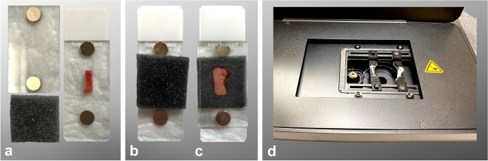
We evaluated 17 tissue samples from mastectomy specimens, rich in adipose tissue, submitted to the department of pathology at the Medical University of Vienna. We conducted our study on the FCM VivaScope® 2500M-G4 (Mavig GmbH, Munich, Germany; Caliber I.D.; Rochester NY, USA).
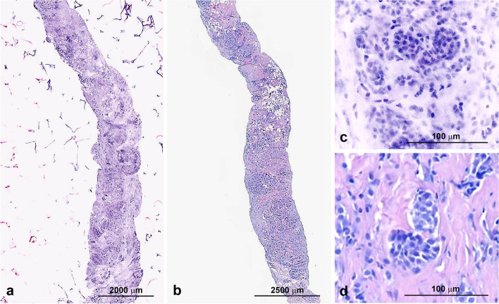
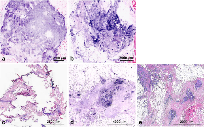
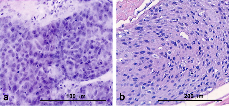
When comparing FCM to frozen sections, we found a very similar overall processing time for FCM images and frozen sections respectively. Image quality was mostly superior to frozen sections (especially for adipose tissue and nuclear detail) but inferior to H&E stained FFPE sections. Limitations of the technology were uneven coloring, invisibility of ink applied for marking tissue margins and distortion artifacts if too much pressure is applied to the tissue.
FCM has the potential to expand the application and usefulness of rapid tissue analysis as speed is comparable and quality exceeds that of frozen sections especially in tissues rich in adipose cells such as breast specimen.
快速的组织学诊断对各种外科手术至关重要。然而,冷冻切片需要专门的实验室设备,容易出现冷冻伪影,并且不适用于所有类型的组织。脂肪组织尤其难以进行冷冻切片处理。尽管这些局限性是众所周知的,但尚未建立任何其他可替代的用于显微镜组织分析的方法来替代冷冻切片。我们的目的是评估基于离体荧光共聚焦显微镜(FCM)的组织成像是否适用于快速评估富含脂肪组织的乳房肿瘤标本。
我们评估了 17 个来自维也纳医科大学病理科的富含脂肪组织的乳房切除术标本的组织样本。我们在 FCM VivaScope® 2500M-G4(Mavig GmbH,慕尼黑,德国;Caliber I.D.;Rochester NY,美国)上进行了研究。
当将 FCM 与冷冻切片进行比较时,我们发现 FCM 图像和冷冻切片的总体处理时间非常相似。图像质量大多优于冷冻切片(特别是对于脂肪组织和核细节),但不如 H&E 染色的 FFPE 切片。该技术的局限性包括颜色不均匀、用于标记组织边界的墨水不可见以及如果对组织施加过多压力会导致失真伪影。
FCM 有可能扩大快速组织分析的应用和实用性,因为其速度相当,并且质量优于冷冻切片,尤其是在富含脂肪细胞的组织中,如乳房标本。